
What is Brugada Syndrome?
Brugada syndrome is a genetic disorder characterized by distinct electrocardiographic abnormalities and an elevated risk of sudden cardiac death resulting from malignant ventricular tachyarrhythmias. The syndrome was named after the Spanish cardiologists, the Brugada brothers (Pedro Brugada, Josep Brugada, and Ramon Brugada), who first described it in 1992 in a cohort of individuals who had suffered sudden death and exhibited a specific electrocardiographic pattern. It was subsequently determined that many of these individuals harbored a genetic defect causing dysfunctional expression of a protein regulating sodium transport (the sodium channel) into the interior of the cardiac cell. The most common and thoroughly studied mutation is localized within the SCN5A gene, although additional causative genes have now been identified. Due to this disruption, and potentially other abnormalities at the cellular level, an ionic imbalance occurs, which can precipitate severe ventricular tachyarrhythmias that cause the heart to beat excessively and ineffectively fast. These ventricular tachyarrhythmias can ultimately prove fatal.
Regarding its epidemiological characteristics, Brugada syndrome is considered rare, with an estimated prevalence of 1–10 per 10,000 individuals in Western societies. It occurs more frequently in males and in Asian populations compared to European cohorts, with a mean age of clinical manifestation at 41 years, although it may present at any age.
Diagnosis
In the majority of cases, patients with Brugada syndrome remain largely asymptomatic. They may, however, experience transient ventricular tachyarrhythmias that cause momentary dizziness or syncope; if these arrhythmias are severe and sustained, they can result in sudden death. These arrhythmias and symptoms frequently manifest during sleep or under conditions of absolute relaxation and rest, driven by autonomic nervous system influence (increased vagal tone).
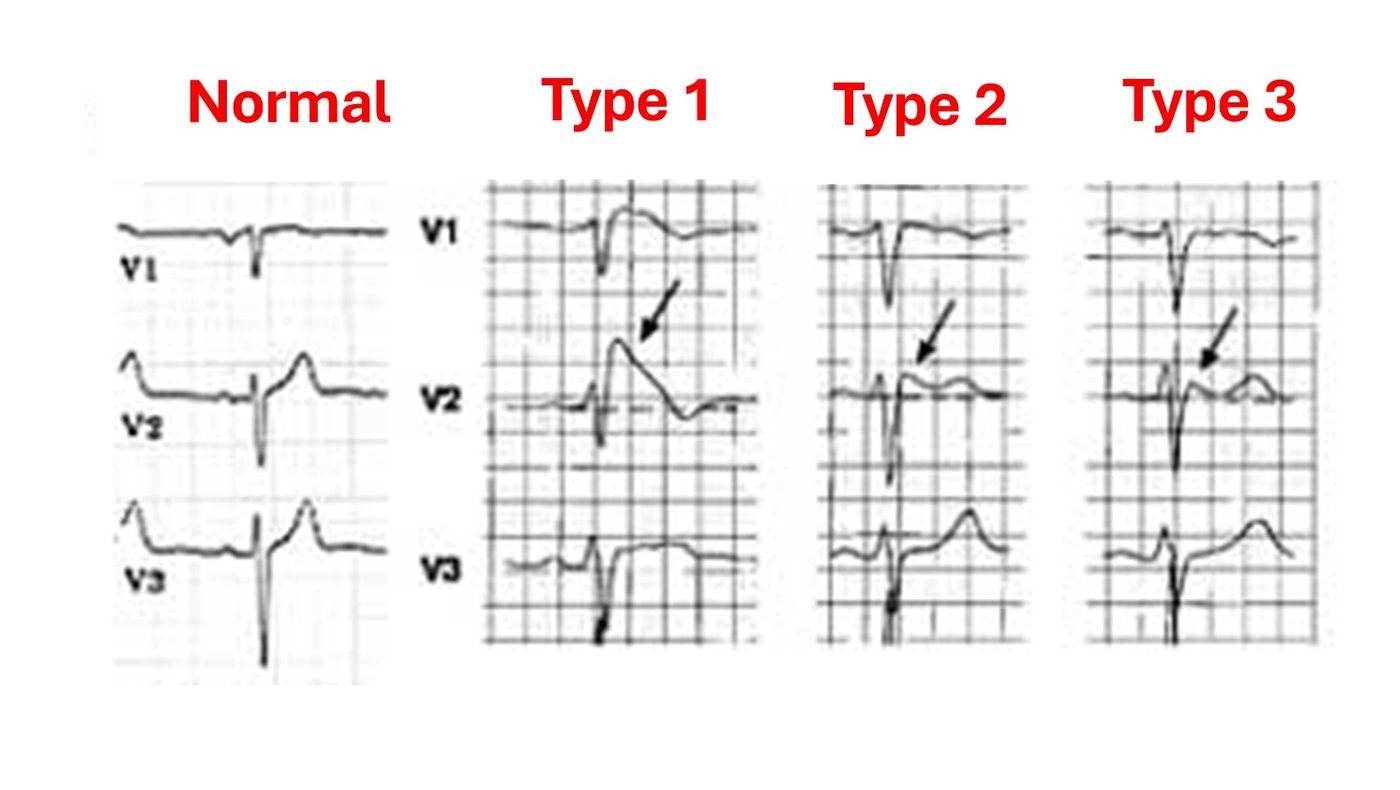
Diagnosis depends fundamentally on identifying characteristic abnormalities on the electrocardiogram. In cases where the electrocardiographic abnormality is present in isolation without clinical symptoms, the condition is classified as a Brugada ECG pattern rather than Brugada syndrome. The diagnosis is frequently confirmed by placing the precordial ECG patches in a higher position, shifted upward by one or two intercostal spaces. Another diagnostic challenge is that the electrocardiographic abnormality can be dynamic, appearing intermittently over time. Occasionally, the Brugada ECG pattern may be unmasked or exacerbated by a high fever, alcohol abuse, or heavy meals, as well as by the use of certain illicit substances (such as cocaine) or the presence of electrolyte imbalances (such as hypokalemia – low blood potassium levels), which can reveal or worsen the clinical picture.
Only the Type 1 ECG pattern is diagnostic of the syndrome, whereas Type 2 and Type 3 patterns strongly raise clinical suspicion but are not diagnostic on their own. A Type 2 or Type 3 pattern can be converted into a Type 1 pattern via an examination known as a pharmacological challenge test, during which an intravenous antiarrhythmic medication (flecainide or procainamide) is administered. Because the syndrome is hereditary, following a definitive diagnosis, proactive clinical and electrocardiographic screening of first-degree relatives (parents, siblings, children) is essential to identify asymptomatic carriers (cascade screening).
Risk Stratification
Patients at the highest risk are those who have already experienced a severe arrhythmic event or have been resuscitated from sudden cardiac death. Furthermore, patients with a prior history of syncope (transient loss of consciousness) are considered to be at high risk for future life-threatening arrhythmic events. Finally, patients who exhibit a spontaneous Type 1 pattern on their electrocardiogram face a greater risk of arrhythmic events compared to patients with a Type 2 or Type 3 pattern.
In contemporary cardiology, risk stratification is performed utilizing specialized multiparametric scoring systems (such as the Shanghai Score), which additionally evaluate a family history of premature sudden death, the presence of a fragmented QRS complex on the ECG, and sinus node dysfunction. Electrophysiological study (EPS) is occasionally utilized as a tool for risk stratification. This is performed in individuals who present with a Brugada ECG pattern but have no documented history of a severe arrhythmic event. The extent to which an electrophysiological study can reliably predict future clinical risk remains a subject of ongoing debate within the scientific community.
Patients with Brugada syndrome do not exhibit structural cardiac dysfunction and, provided they are protected from the risk of a severe ventricular arrhythmia, they can lead an entirely normal life.
Management
Conservative & Preventive Measures (Applicable to all patients):
- Strict avoidance of medications that can exacerbate the underlying abnormalities of the syndrome (detailed listings are available on the official website: www.brugadadrugs.org).
- Prevention of hyperpyrexia (high fever) through the immediate administration of antipyretics.
- Avoidance of alcohol abuse and excessively heavy meals.
- Rigorous maintenance of systemic electrolytes within normal physiological ranges and the absolute avoidance of illicit, addictive substances.
Pharmacological Therapy:
Data supporting the management of Brugada syndrome with long-term pharmacological therapy remain limited. Among antiarrhythmic agents, quinidine is the only medication that may be indicated for prophylactic therapy. Additionally, isoproterenol, which is administered exclusively via the intravenous route, serves as a useful inpatient medication to suppress an acute, severe storm of arrhythmic episodes.
Invasive Therapies (For high-risk patients):
- Implantable Cardioverter-Defibrillator (ICD) Placement: Once a patient is stratified as high-risk, the only established, definitive therapy is the implantation of a cardioverter-defibrillator.
- Catheter Ablation: In patients experiencing frequent arrhythmias, recurrent shocks from the defibrillator, or electrical storms, epicardial catheter ablation is now performed with high success rates. This invasive procedure is carried out on the outer surface of the heart (specifically the right ventricular outflow tract) and effectively eliminates the underlying arrhythmogenic substrate.