Biventricular pacing, or Cardiac Resynchronization Therapy (CRT), is an onboarding, proven effective treatment for specific categories of patients suffering from heart failure.
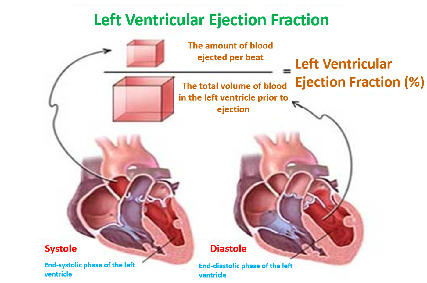
Millions of people worldwide suffer from congestive heart failure. This is a condition where the heart muscle is weakened, making it difficult for the heart to effectively pump blood and meet the body’s metabolic demands. In these patients, the left ventricular ejection fraction is low, specifically under 35-40%. The heart’s inability to function properly leads to lower extremity edema, shortness of breath (dyspnea), and reduced exercise tolerance, even during mild physical activity.
Cardiac Dyssynchrony
One of the primary factors contributing to this diminished cardiac output in heart failure is cardiac dyssynchrony. This means that, in addition to the structural weakness of the myocardium, the sequence in which the various chambers and regions of the heart contract is disrupted, lacking the precise coordination seen in a normal heart that ensures seamless function. This lack of synchronization can occur between the atria and the ventricles (atrioventricular dyssynchrony), between the right and left ventricles (interventricular dyssynchrony), or within the left ventricle itself (intraventricular dyssynchrony).
In intraventricular dyssynchrony, specific regions of the left ventricle that are closer to the cardiac conduction system (the network of specialized electrical pathways that transmit impulses to trigger contraction)—such as the interventricular septum—contract earlier than more remote regions, like the lateral wall. Even when this mechanical delay is measured in just a few thousandths of a second (40-80 milliseconds), it is significant enough to severely compromise overall cardiac output.
Intraventricular dyssynchrony can be visualized and measured via a standard echocardiogram. However, its most definitive indication is provided by a simple electrocardiogram (ECG) showing a Left Bundle Branch Block (LBBB). This condition signifies a conduction block in the electrical pathway that delivers impulses to the left ventricle.
Cardiac Resynchronization Therapy / Biventricular Pacing
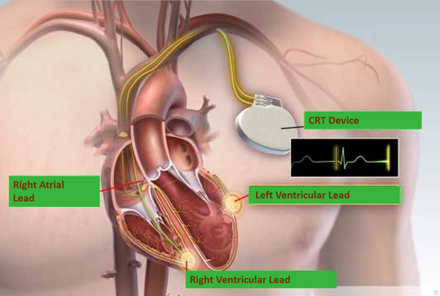
Biventricular pacing aims to restore the heart’s lost mechanical coordination by targeting and correcting all three types of cardiac dyssynchrony mentioned above. In cardiac resynchronization systems, in addition to the two standard leads placed in the right atrium and right ventricle—which are typically used in conventional pacemakers or implantable cardioverter-defibrillators (ICDs)—a third lead is implanted. This left ventricular lead is introduced through the venous system of the coronary sinus to pace the epicardial (outer) surface of the left ventricle.
When the right and left ventricular leads deliver electrical impulses simultaneously (biventricular pacing), ventricular dyssynchrony (both inter- and intraventricular) is corrected. Furthermore, when the system coordinates atrial contraction with ventricular contraction, atrioventricular synchrony is achieved.
Consequently, biventricular systems correct one of the most critical pathophysiological causes of heart failure, thereby increasing cardiac output and improving patient symptoms. These systems can be configured either as a standalone pacemaker (CRT-P) or combined with an implantable cardioverter-defibrillator (CRT-D). In the latter case, besides the therapeutic benefits of resynchronization, the device provides immediate intervention and therapy if a life-threatening ventricular arrhythmia occurs. Numerous clinical trials have demonstrated that heart failure patients managed with CRT devices experience a statistically significant improvement in overall survival and life expectancy.
Surgical Procedure
The implantation procedure is performed in a manner highly similar to that of a conventional pacemaker. Typically, the left pectoral region is preferred. Under local anesthesia, a 5-centimeter incision is made inferior to the clavicle. The three leads are introduced through either the subclavian or the cephalic vein, advanced into their precise positions within the cardiac chambers, and secured to the underlying pectoral muscle with sutures.
The leads are then connected to the pulse generator, which is placed subcutaneously into a specially prepared pocket. Finally, the incision is sutured, and the patient is transferred back to the regular ward, as intensive care admission is not required. Patient mobilization occurs on the same or the following day, and the procedure generally requires only a single overnight hospital stay. Mild localized discomfort may be present during the first few postoperative days, which is well-managed with standard over-the-counter analgesics.
Complications
The complications associated with the implantation of a biventricular system (CRT-P or CRT-D) are largely identical to those encountered in standard pacemaker or ICD implantations.
Adverse events specific to cardiac resynchronization systems include:
- Lead dislodgement: This typically involves the left ventricular lead (occurring in less than 3% of cases in high-volume, experienced centers). Dislodgement can lead to a loss of cardiac resynchronization, resulting in clinical decompensation and the recurrence of heart failure symptoms.
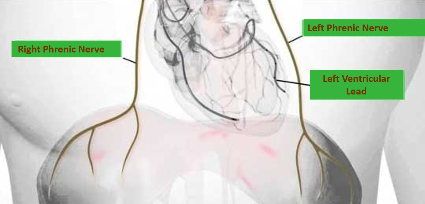
- Phrenic nerve stimulation: The close anatomical proximity of the left phrenic nerve (which innervates the left hemidiaphragm) to the left ventricular lead can cause diaphragmatic stimulation. This presents as involuntary, rapid diaphragmatic contractions synchronized with every heartbeat.
In almost all cases, phrenic nerve stimulation can be resolved non-invasively via electronic device reprogramming. Adjusting the pacing vectors or parameters eliminates phrenic nerve recruitment while maintaining effective left ventricular pacing.
 Follow-up Care for Patients with Biventricular Systems
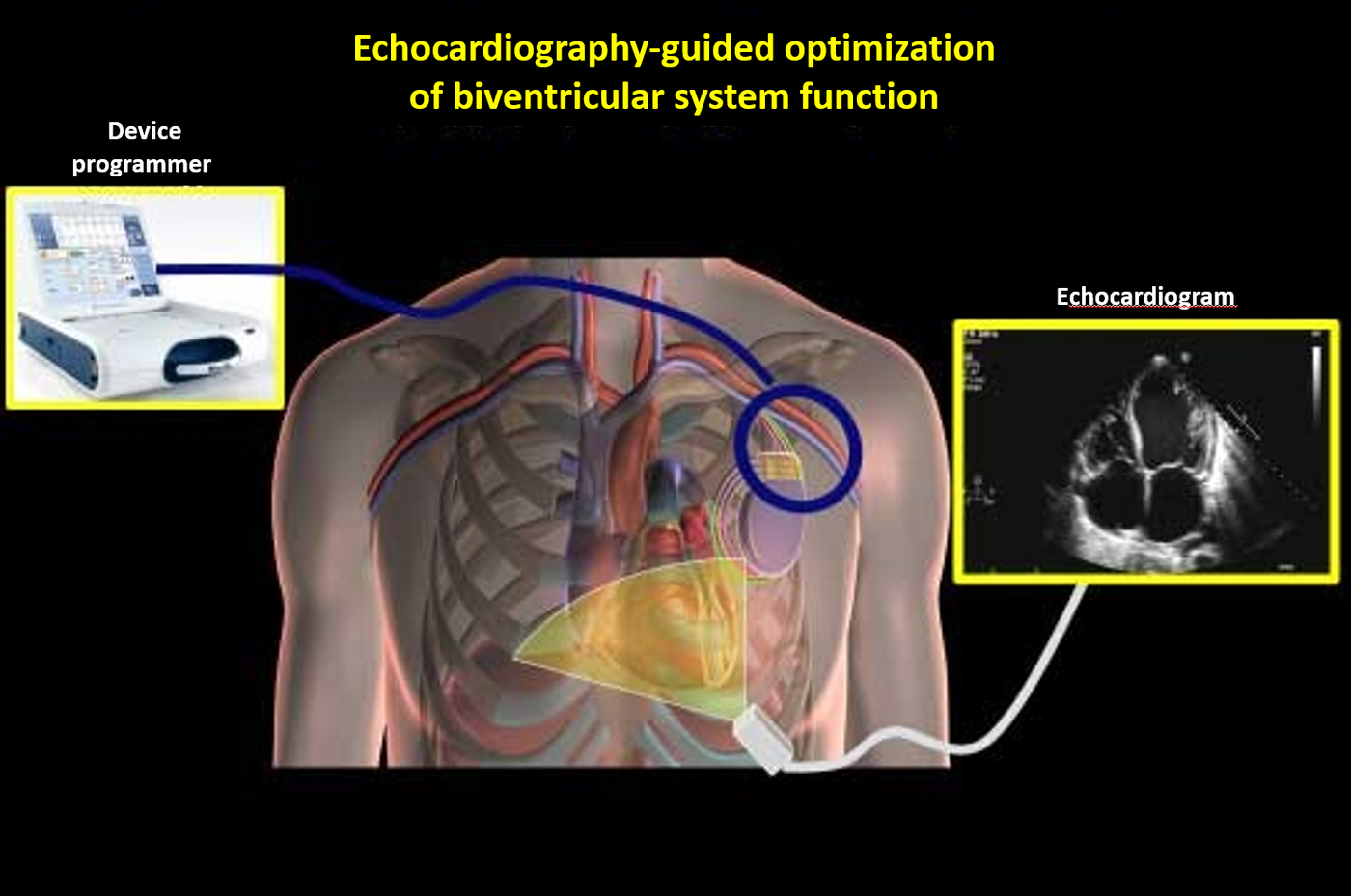
Biventricular systems, like all pacemakers and defibrillators, are monitored non-invasively using dedicated medical computers known as device programmers, which are available at specialized implanting centers. Each device manufacturer utilizes a proprietary programmer. These programmers allow clinicians to evaluate system functionality, retrieve diagnostic data, and optimize device settings. The initial post-implantation evaluation is typically scheduled at one month, followed by routine follow-up assessments every six months.
Occasionally, particularly when patients do not exhibit the anticipated clinical response post-implantation, a formal CRT optimization procedure is performed. This process is guided by transthoracic echocardiography and electrocardiography (ECG). By non-invasively adjusting the device parameters via the programmer during real-time imaging, clinicians can identify the precise settings that yield optimal hemodynamic and cardiac function.