
(For further details regarding this arrhythmia, please refer to Arrhythmias → Tachyarrhythmias → Atrioventricular Nodal Reentrant Tachycardia – AVNRT)
Indications for Catheter Ablation
Catheter ablation for AVNRT is a minimally invasive procedure that provides a definitive therapeutic solution. Ablation for this specific type of tachycardia represents one of the few interventions in cardiology capable of achieving a permanent cure, eliminating the need for subsequent pharmacological or other long-term management.
Candidates for catheter ablation include:
- Patients with a documented episode of supraventricular tachycardia (specifically, atrioventricular nodal reentrant tachycardia).
- Patients who fail to achieve adequate arrhythmia control with pharmacological therapy and continue to experience symptomatic episodes.
- Patients who must discontinue antiarrhythmic therapy due to severe adverse effects.
For instance, patients developing bradycardia must discontinue relevant medications. In such cases, recommending permanent pacemaker implantation solely to sustain antiarrhythmic drug compliance is an inappropriate clinical approach. Catheter ablation can resolve the underlying arrhythmia, rendering subsequent pharmacological therapy unnecessary. - Patients who prefer to avoid long-term pharmacological prophylaxis.
Although recurrent episodes can be mitigated to some extent via pharmacotherapy, the agents typically prescribed (such as beta-blockers or calcium channel blockers) predominantly induce bradycardia and systemic symptoms, including fatigue and lethargy. Consequently, these medications may be poorly tolerated, particularly by young and physically active individuals. - Patients residing in remote areas where access to immediate hospital care during an acute tachycardic episode is limited.
- Young women planning a pregnancy, given that the onset or recurrence of this specific tachycardia during gestation presents significant management challenges.
Pharmacological interventions, whether administered for acute termination or long-term prophylaxis, carry inherent risks of fetal toxicity. Furthermore, performing catheter ablation during pregnancy is contraindicated due to mandatory fluoroscopic radiation exposure. Although non-fluoroscopic, zero-fluoroscopy mapping techniques exist, they are generally avoided during pregnancy because absolute safety regarding the technical performance of the procedure itself cannot be fully guaranteed.
Procedural Objective
As detailed in the pathophysiological description of the arrhythmia, AVNRT is mediated by a micro-reentrant circuit located within and adjacent to the atrioventricular (AV) node. In the vast majority of cases, this circuit involves two distinct pathways: the fast pathway and the slow pathway. The therapeutic objective of catheter ablation is the targeted modification or ablation of the slow pathway, which disrupts the reentrant mechanism and prevents tachycardic recurrence. Typically, one or two radiofrequency energy applications, lasting approximately one minute each, are sufficient to achieve this endpoint. Following therapeutic delivery, programmed electrical stimulation is performed in an attempt to reinduce the tachycardia. Intravenous isoproterenol may also be administered to facilitate tachycardic induction. The non-inducibility of the arrhythmia post-ablation serves as the primary endpoint of procedural success.
Pre-procedural Patient Preparation
The ablation procedure does not require extensive pre-operative preparation. General guidelines include the following:
- Discontinuation of all antiarrhythmic agents for at least 5 days prior to the procedure.
- If the patient is receiving amiodarone, a significantly longer washout period is required (>20 days).
Patients who fear that drug discontinuation may trigger an arrhythmia recurrence during this interval should be reassured. Performing the procedure under the effect of antiarrhythmic medications can suppress arrhythmia inducibility, lead to an incorrect electrophysiological diagnosis, mask residual pathway conduction, and ultimately result in procedural failure for an otherwise highly effective intervention. - Anticoagulants or other antithrombotic therapies typically do not require discontinuation; however, explicit prior consultation with the attending physician is mandatory.
- Other concurrent pharmacological therapies may be continued normally, including on the morning of the procedure.
- The patient must fast on the morning of the procedure (a standard evening meal the night before is permissible). If the procedure is scheduled for the afternoon, a light breakfast may be consumed.
- Upon hospital admission, the patient must provide all previous medical records, with particular emphasis on prior electrocardiograms (ECGs).
Electrophysiological Procedure
The procedure is typically performed under local anesthesia at the vascular access sites, without the need for general anesthesia. Conscious sedation may be administered to highly anxious patients, though it is generally not recommended.
In the electrophysiology (EP) laboratory, the patient is placed under continuous electrocardiographic monitoring. Following local anesthetic infiltration, percutaneous punctures are performed in the femoral region to introduce vascular sheaths into the femoral veins. Electrophysiological catheters (diagnostic electrode catheters) are advanced through these sheaths and positioned at specific target sites within the cardiac chambers to record intracardiac electrograms. These electrical signals are displayed on the laboratory’s multi-channel system and mapping systems. An electrophysiological study follows, involving programmed ventricular and atrial stimulation to induce the arrhythmia, establish the precise mechanism, and confirm the diagnosis. Targeted ablation is then performed using a specialized steerable therapeutic catheter (ablation catheter). Once the ablation catheter is optimally positioned at the target site, radiofrequency energy is delivered. Post-ablation testing is subsequently conducted to confirm arrhythmia non-inducibility. A mandatory observation period of 20 to 30 minutes is maintained, during which intravenous isoproterenol is infused to challenge the ablation line. If the tachycardia remains completely non-inducible, the procedure is deemed successful. Hemostasis is achieved at the venous puncture sites, and the patient is transferred to the recovery area briefly before returning to the inpatient ward.
Post-procedural Management and Recovery
Immediately following the procedure, the patient returns to the ward and remains on strict bed rest, ensuring that the ipsilateral lower extremity is kept immobilized during the initial hours. Patients are typically ambulated 4 to 5 hours post-procedure. A standard 12-lead ECG is performed the following morning, and the patient is discharged after receiving specific post-operative instructions. Routine professional and physical activities can be resumed two days after the intervention. Since arterial access is rarely required for AVNRT ablation (femoral arterial puncture is exceptionally uncommon), the patient may return to vigorous physical exertion, gym workouts, and sports activities as early as the second post-operative day.
Expected Outcomes and Efficacy
The primary clinical objective of the procedure is permanent eradication of the arrhythmia and long-term freedom from recurrence. The acute procedural success rate exceeds 95%. In highly uncommon instances, despite meeting successful intraoperative endpoints, a late recurrence of the arrhythmia may occur in approximately 2% to 5% of cases. In these recurrent scenarios, a repeat ablation procedure can be safely performed.
Procedural Risks and Potential Complications
When performed in high-volume, experienced electrophysiology laboratories, catheter ablation for AVNRT is an exceedingly safe intervention, with an overall complication rate below 1%. The most significant procedural complication is the induction of iatrogenic atrioventricular (AV) block, resulting from permanent injury to the AV node. In historical published series—which include data from the early developmental eras of catheter ablation—the reported incidence of permanent AV block is approximately 0.5%. This risk arises when the slow pathway or reentrant circuit resides in close anatomical proximity to the compact AV node. However, contemporary electrophysiological mapping and titration techniques render this complication virtually negligible in experienced centers. Within our clinical team, spanning more than 20 years of experience and over 1,200 AVNRT ablation procedures, this complication has never occurred.
Other potential complications inherent to any cardiac catheterization and ablation procedure include:
- Access-site hematoma at the vascular puncture locations.
- Cardiac tamponade.
Cardiac tamponade involves the escape of blood from the cardiac chambers into the pericardial space (the sac surrounding the heart). This is an extremely rare complication during AVNRT ablation. It constitutes a medical emergency requiring immediate management via percutaneous pericardiocentesis to drain the accumulated blood, which is typically curative. A micro-drainage catheter is maintained in the pericardial space for approximately 24 hours under close hemodynamic monitoring and is subsequently removed. This complication generally extends the hospital stay by 2 to 4 days. - Pneumothorax or hemothorax (secondary to subclavian vein puncture).
Subclavian vein access is routinely avoided during catheter ablation for AVNRT.
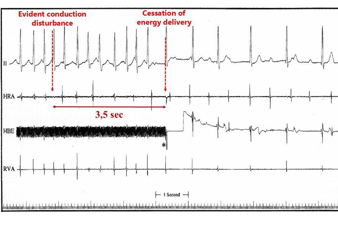
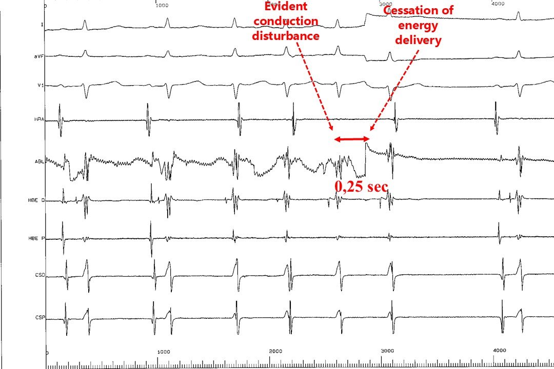 Figure 1. During the delivery of radiofrequency energy for the treatment of AVNRT, a distinct accelerated junctional rhythm (junctional ectopy) typically emerges from the atrioventricular node. It is imperative to meticulously monitor ventriculoatrial (VA) and anterograde conduction during this rhythm. Impaired retrograde conduction to the atria may indicate impending injury to the compact AV node and necessitates the immediate cessation of energy delivery.
The upper tracing (reproduced from a textbook of cardiology) demonstrates a case where energy delivery was continued for approximately 3.5 seconds despite apparent conduction impairment. This resulted in permanent, irreversible AV nodal injury requiring permanent pacemaker implantation.
The lower tracing (from our electrophysiology laboratory) illustrates that energy delivery is terminated within 0.25 seconds of detecting a similar conduction disturbance. Vigilant real-time monitoring and rapid reaction times successfully avert this highly undesirable adverse outcome.