
(For further information regarding the arrhythmia, see Arrhythmias → Tachyarrhythmias → Atrial Flutter)
As previously delineated, atrial flutter is classified into typical, which involves a macro-reentrant circuit within the right atrium passing through the zone between the inferior vena cava and the tricuspid valve annulus (cavotricuspid isthmus – CTI), and atypical, which encompasses any atrial tachycardia where the macro-reentrant circuit is localized in other anatomical regions.
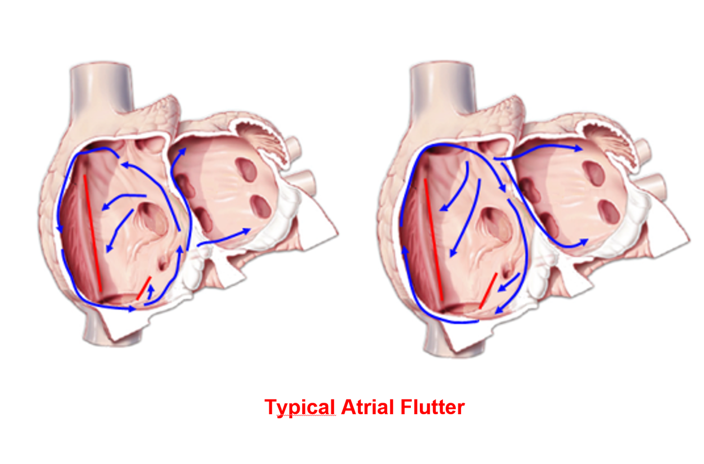
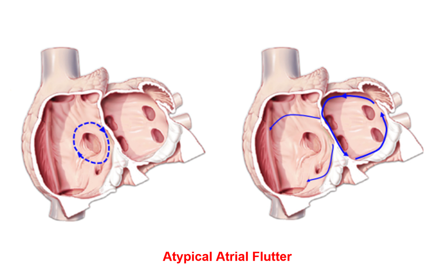
Indications for Ablation
Ablation of typical atrial flutter is a minimally invasive technique that offers a definitive therapeutic cure. Candidates for the procedure include:
- Patients with a documented history of a symptomatic episode of typical atrial flutter, as identified via electrocardiogram (ECG).
- Patients who are refractory to antiarrhythmic drug therapy and exhibit persistent arrhythmia.
- Patients in whom pharmacological rate control cannot be adequately achieved.
- Patients who develop severe adverse effects from medical therapy, necessitating its discontinuation.
For instance, patients presenting with bradycardia must discontinue rate-limiting or antiarrhythmic medications. The alternative of permanent pacemaker implantation solely to tolerate medical therapy—though occasionally recommended—is clinically inappropriate. Catheter ablation successfully resolves the arrhythmia, eliminating the need for long-term pharmacotherapy.
Procedural Target
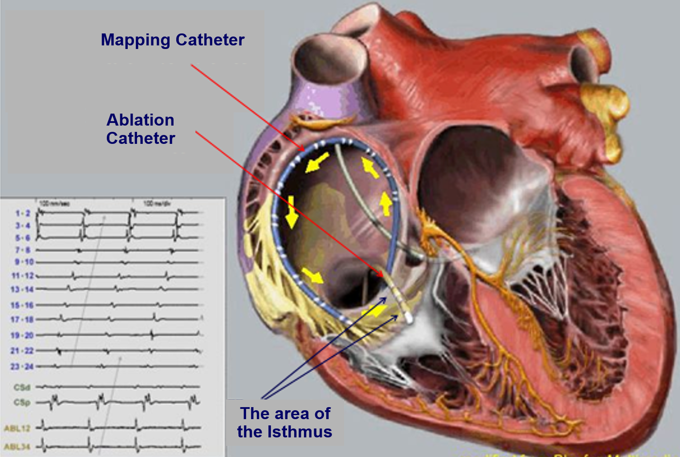
The electrophysiological objective in typical, isthmus-dependent atrial flutter is the creation of a linear lesion across the cavotricuspid isthmus, spanning the narrowest region from the tricuspid valve annulus to the inferior vena cava. This acute endpoint is achieved in approximately 100% of cases. Following lesion deployment, differential pacing from both sides of the ablation line must be performed to confirm the presence of a complete, bidirectional conduction block. After a brief observation period, the patient is discharged from the electrophysiology laboratory.
Pre-Procedural Patient Preparation
The ablation procedure requires minimal specific preparation by the patient. The standard protocol includes:
- If the patient is on anticoagulant or other antithrombotic therapy, cessation is generally not required; however, management must be pre-coordinated with the treating physician.
- All other chronic medications may be continued normally, including on the morning of the procedure.
- The patient must maintain NPO status (fasting) from the morning of the procedure (a standard evening meal the night before is permissible). If the procedure is scheduled for the afternoon, a light breakfast may be consumed.
- Upon hospital admission, previous diagnostic records, particularly prior electrocardiograms, must be available.
The Ablation Procedure
Most commonly, the procedure is performed without general anesthesia, utilizing local anesthesia strictly at the venous access sites. If clinically indicated, deeper conscious sedation can be administered.
In the electrophysiology laboratory, the patient is placed under continuous ECG monitoring. Under local anesthesia, percutaneous punctures are performed in the femoral region to introduce specialized vascular sheaths into the femoral veins. Through these sheaths, electrophysiology catheters (multipolar electrode catheters) are advanced and positioned at specific sites within the cardiac cavities. These catheters record intracardiac electrograms, which are displayed on the laboratory’s multi-channel recording and electroanatomical mapping systems.
An electrophysiological study follows, which may involve programmed atrial stimulation to induce the arrhythmia. However, induction is often unnecessary because the arrhythmia is either already present or its diagnosis has been clearly established by a baseline 12-lead ECG during sinus rhythm.
If the arrhythmia is ongoing or induced, the diagnosis is confirmed, followed by targeted ablation using a specialized, steerable ablation catheter. Once stable contact is achieved at the target site, radiofrequency (RF) energy is delivered. Sequential, point-by-point lesions are created across the isthmus to construct a continuous line of conduction block. Subsequently, pacing maneuvers are performed on either side of the line to verify a complete, gap-free bidirectional block.
If the electrophysiological evaluation reveals a circuit that does not involve the cavotricuspid isthmus (atypical atrial flutter), 3D electroanatomical mapping is utilized to reconstruct the activation sequence, localize the critical isthmus, and guide targeted ablation.
A standard waiting period of 20 to 30 minutes is observed to ensure lesion durability. If block persists, the procedure is deemed successful and concluded. Hemostasis at the femoral puncture sites is achieved, and the patient is transferred to the ward, often following a brief stay in the recovery area.
Post-Procedural Course
Immediately post-procedure, the patient returns to the ward where they remain at strict bed rest, avoiding vigorous movement of the lower extremities during the initial hours. Ambulation is typically permitted 4 to 5 hours post-recovery. On the following morning, a standard ECG is performed, and after receiving discharge instructions, the patient is allowed to leave the hospital. Normal occupational and physical activities can be resumed on the second post-operative day.
Temporary anticoagulant therapy is usually required for a designated period, in accordance with the specific instructions provided by the attending cardiologists.
Expected Outcomes
The primary clinical objective is the permanent restoration and maintenance of sinus rhythm. This goal is highly achievable in patients without a history of atrial fibrillation (AF). Conversely, patients with a pre-existing history of comorbid AF carry a higher risk of presenting with recurrent AF episodes post-ablation, despite the definitive eradication of the typical atrial flutter circuit. The success rate for eliminating typical atrial flutter approaches 100%, while the risk of major clinical complications remains negligible.
Procedural Risks and Complications
In high-volume, experienced centers, typical atrial flutter ablation is an exceedingly safe procedure. Even in elderly populations with multiple comorbidities, the overall complication rate remains below 1%.
Potential complications, inherent to any catheter ablation procedure, include:
- Hematoma at the vascular access sites.
- Cardiac Tamponade: This involves the extravasation of blood into the pericardial space, leading to accumulation between the epicardium and the pericardial sac. Cardiac tamponade is an extremely rare complication during this specific procedure. It represents a medical emergency requiring immediate management via pericardiocentesis to drain the accumulated blood, which is typically curative. A small indwelling drainage catheter remains in the pericardial space for approximately 24 hours prior to removal, during which time the patient undergoes intensive monitoring. This complication generally extends the hospital stay by 2 to 4 days.
- Pneumothorax or Hemothorax: This risk is associated strictly with subclavian vein puncture. However, subclavian venous access is not routinely performed during standard atrial flutter ablation procedures.