Ablation of Accessory Pathways
(For further details, see Arrhythmias → Tachyarrhythmias → Tachycardias involving an accessory pathway – WPW syndrome)
The diagnosis of an accessory pathway is typically established during childhood via an initial electrocardiogram. As outlined in the section describing arrhythmias, once an accessory pathway is diagnosed, a diagnostic evaluation must be performed to assess the current risk of a major arrhythmic event. In other cases, where no prior electrocardiogram has been performed, the diagnosis is made immediately after the patient experiences a symptomatic episode of tachycardia. Naturally, concealed pathways also exist, where the diagnosis is made following the clinical manifestation of tachycardia, with the definitive diagnosis established via an electrophysiological study.
In which patients is ablation indicated?
Ablation of the causative accessory pathway terminates episodes of tachycardia and eliminates the risk of future severe arrhythmic events. It is a minimally invasive technique that provides a definitive solution to the problem. Like very few therapies, not only in cardiology but in medicine in general, it achieves a permanent cure without the need for subsequent pharmaceutical or other medical maintenance.
The following patient categories are candidates for ablation:
- Patients with manifest pre-excitation and a history of a severe arrhythmic event (e.g., atrial fibrillation with ultra-rapid conduction over the pathway).
- Patients with manifest pre-excitation and episodes of tachycardia mediated by the pathway, regardless of the pathway’s inherent risk profile.
- Patients with episodes of tachycardia, even without manifest pre-excitation (concealed pathways), with the objective of eliminating these episodes.
- Asymptomatic patients with a high-risk pathway, as demonstrated by an electrophysiological study. Even if the pathway is proven to be of low risk for major arrhythmic events (without ruling out future benign episodes of tachycardia) and the patient has already undergone an electrophysiological study, ablation should ideally be attempted during the same procedure, provided that the operating physician possesses the experience and capability to achieve ablation rapidly and safely.
- Patients who have initially tried and are not adequately controlled by antiarrhythmic medication, continuing to present with episodes of tachycardia.
- Patients in whom pharmaceutical therapy caused a serious adverse effect and must therefore be discontinued.
- Patients with an accessory pathway, even if asymptomatic, who work in high-responsibility occupations (e.g., pilots, bus drivers).
- Athletes with an accessory pathway.
- Young women planning a pregnancy, given that the onset of tachyarrhythmias during gestation can complicate therapeutic management. The administration of pharmaceuticals, both for terminating acute episodes and for prophylaxis, can never be considered entirely safe for the fetus. Additionally, performing an ablation procedure during pregnancy is contraindicated due to radiation exposure from cardiac fluoroscopy. Although non-fluoroscopic mapping approaches exist, they are not preferred as they cannot guarantee absolute safety during the execution of the procedure itself.
Ablation is naturally the first-line therapy for accessory pathways. However, there are cases where conservative management is preferable:
- Young age of the patient.
- Young age of the patient combined with a pathway location in close proximity to the normal conduction system.
- Small body habitus of the young patient.
If, despite these factors, indicators of high risk or already manifested severe symptoms are present, ablation can be attempted even at a young age. Furthermore, regarding the anatomic location of the pathway, in experienced hands, ablation remains safe even if the pathway is located very close to the normal conduction system.
What is the procedural objective?
The primary objective of the procedure is the complete destruction of the pathway, following an initial diagnostic evaluation that reveals its involvement in arrhythmias and its risk profile. The pathway must be eliminated via the delivery of radiofrequency energy and not through mechanical trauma. This is because, during the procedure, there is a small probability that the pathway may be transiently injured by the catheters and thus become angiographically or electrically hidden, preventing targeted ablation, only to recur in the future. Following energy delivery, testing is performed to confirm full (orthodromic and antidromic) elimination of the pathway, followed by a waiting period (approximately 30 minutes) to ensure the stability of the successful result.
Patient preparation for the procedure
The ablation procedure does not require extensive preparation by the patient. Generally, it requires:
- Discontinuation of all antiarrhythmic medications for at least 5 days prior to the procedure.
- If the medication is amiodarone, a significantly longer discontinuation period (>20 days) is mandatory.
Patients who fear that discontinuing their medication might trigger an episode in the interim should be reassured. Performing the procedure under the influence of antiarrhythmic drugs can completely conceal the pathway and prevent the induction of tachycardias, leading to an incorrect procedural diagnosis, misleading outcomes, and overall procedural failure of an otherwise highly effective intervention.
- If the patient receives antithrombotic medication, management must be coordinated with the physician regarding the periprocedural administration protocol.
- Regarding other chronic pharmaceutical treatments, these may be continued normally, even on the day of the procedure.
- The patient must remain fasting on the morning of the procedure (a normal evening meal the night before is permissible), whereas if the procedure is scheduled for the afternoon, a light breakfast may be consumed.
- Upon admission to the hospital, the patient must bring previous diagnostic examinations, particularly prior electrocardiograms.
The procedural technique
Most commonly, the procedure is performed without general anesthesia, utilizing only local anesthesia at the puncture sites. However, for young or highly anxious patients, deeper sedation is administered.
In the electrophysiology laboratory, the patient is connected to continuous electrocardiographic monitoring. Under local anesthesia, punctures are performed in the femoral region to insert specialized vascular sheaths into the femoral veins. Through these sheaths, electrophysiological catheters (small electrodes) are advanced and positioned at specific sites within the cardiac cavities. These catheters record electrical signals from the cardiac chambers, which are displayed on the laboratory’s mapping systems.
A three-dimensional electroanatomical mapping system may also be utilized, allowing real-time visualization and navigation of the catheters without the use of fluoroscopy. This is followed by the electrophysiological study, which includes assessing the pathway’s risk profile and attempting arrhythmia induction.
Subsequently, ablation therapy is performed using a specialized steerable catheter, the ablation catheter. Depending on the anatomical position of the pathway, the ablation catheter is introduced either via the femoral vein or the femoral artery. In rare cases of anteroseptal pathways, the subclavian vein may be utilized. Detailed mapping is performed with the ablation catheter to localize the exact position of the pathway. Once identified, radiofrequency energy is delivered to achieve immediate destruction of the pathway.
Immediately after, re-testing is conducted to fully confirm the eradication of the pathway. Typically, a 30-minute waiting period is required, and if the successful result persists, the procedure is deemed successful and concluded. Hemostasis is achieved at the puncture sites, and the patient, after spending a brief period in the recovery area, returns to the ward.
Post-procedural care
Immediately after the procedure, the patient returns to the ward, where they remain strictly supine, taking care during the first few hours to avoid intense movements of the lower extremities. Typically, the patient can ambulate 4–5 hours after returning to the ward, provided only venous punctures were performed. Mobilization may be deferred, up to the following morning at most, if an arterial puncture was required. The next morning, an electrocardiogram is performed, and the patient is discharged from the hospital after receiving specific instructions. On the second post-operative day, the patient can safely return to work and resume normal daily activities. If a femoral artery puncture was performed, the patient must avoid strenuous physical activity (sports, gym, etc.) for 4–5 days. If no arterial puncture was performed, return to strenuous activities can occur on the second post-operative day.
What is the expected outcome of ablation?
The expected outcome of the procedure is the complete eradication of the accessory pathway. The success rate exceeds 90%. The probability of recurrence with reappearance of the pathway is less than 5%.
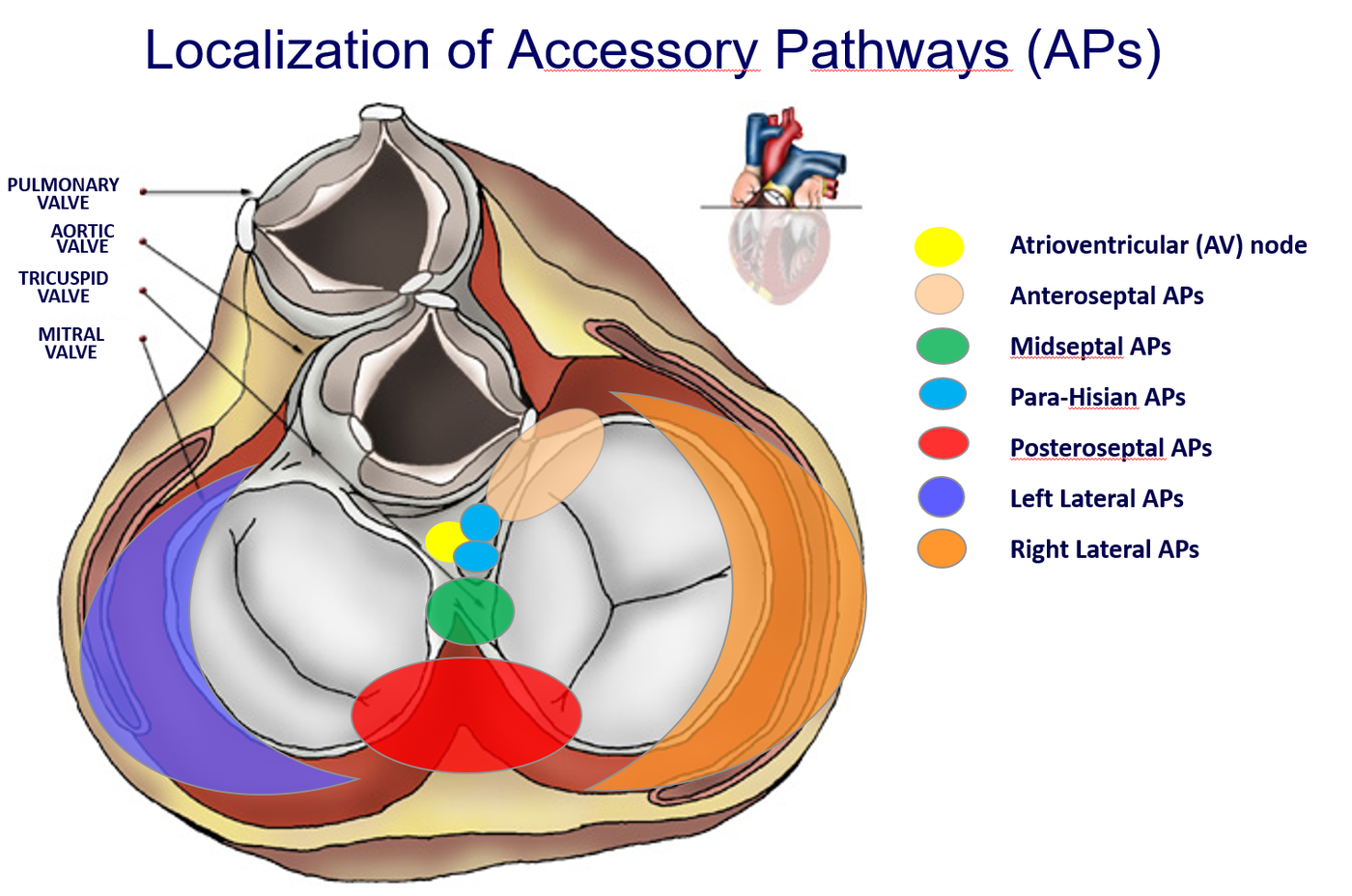
An exception is generally noted for pathways located close to the atrioventricular (AV) node (anteroseptal, midseptal, and particularly para-Hisian pathways), where lower initial energy levels are sometimes delivered due to the risk of inducing permanent damage to the AV node, resulting in a recurrence rate of approximately 10%. If the pathway recurs, this typically manifests within the first few days following ablation. In rare cases, while the pathway remains manifestless on the ECG, tachycardia recurs. In such instances, orthodromic conduction is likely eliminated while retrograde conduction through the pathway persists. If pathway reappearance or tachycardia recurrence occurs, the procedure can be repeated.
Are there risks associated with the procedure?
Accessory pathway ablation is a highly safe procedure when performed in experienced laboratories. The complication rate is typically below 1%. The primary complications include:
- Hematoma at the vascular access sites.
- Damage to the atrioventricular node causing atrioventricular block, necessitating permanent pacemaker implantation. This risk is non-existent for right and left lateral wall pathways, as well as for posteroseptal pathways. Furthermore, with appropriate expertise, ablation of pathways located near the AV node (anteroseptal, midseptal, and para-Hisian pathways) is safe, with a risk profile below 0.3%–0.5%. In these scenarios, if deemed appropriate by the clinical team for enhanced safety, cryo-ablation may be utilized.
- Cardiac tamponade. Cardiac tamponade is the escape of blood from the heart and its accumulation between the outer wall of the myocardium and the pericardium (the sac surrounding the heart). Tamponade is an extremely rare complication in this specific procedure. It constitutes a medical emergency that requires immediate management via pericardiocentesis to drain the accumulated blood. Usually, this intervention is definitive. A microscopic drainage catheter remains in the pericardial space for approximately 24 hours and is subsequently removed while the patient is monitored closely. Typically, a tamponade complication necessitates an extension of hospitalization by 2–4 days.
- Pneumothorax or hemothorax (if subclavian vein puncture is performed). This approach is generally avoided but may be deemed necessary during the ablation of anteroseptal pathways to ensure greater catheter stability.
- Damage to the aortic valve resulting from catheter manipulation.