
What is Sudden Cardiac Death?
Sudden cardiac death (SCD) is defined as an unexpected death occurring within 1 hour of symptom onset (if witnessed), or within 24 hours of the individual last being seen symptom-free (if unwitnessed), in the absence of an obvious non-cardiac cause of death.
Despite significant scientific advancements in recent years, sudden cardiac death remains among the leading causes of mortality in Western societies, accounting for 50% of all fatal cardiovascular events. The socio-economic burden is overwhelming, considering that a large percentage of those affected are within their most productive years of life.
Sudden death stems from an abrupt inability of the heart to meet its workload, preventing vital organs—primarily the brain—from receiving the oxygenated blood they require. Without immediate medical intervention to resolve the cardiac issue (e.g., termination of a severe arrhythmia), death inevitably ensues within minutes. For terminological clarity, a distinction must be made between sudden cardiac death—frequently referred to as cardiac arrest—and syncope. In syncope, the cardiac underlying issue causing transient loss of consciousness due to cerebral hypoperfusion resolves spontaneously, leading to an automatic recovery of consciousness.
What are the prodromal symptoms?
In approximately half of all cases, no warning symptoms are reported. However, chest pain or discomfort may precede the event if an acute cardiac episode is unfolding (e.g., acute myocardial infarction, aortic dissection). Additionally, episodes of palpitations accompanied by dizziness, as well as brief periods of loss of consciousness, may occur. Generally, what warrants particular concern and should prompt immediate medical evaluation is the onset of novel, unprecedented symptoms which, even if transient, do not constitute a familiar daily sensation.
What are the causes?
The immediate cause of sudden cardiac death is a severe cardiac rhythm disorder (arrhythmia) that causes the heart to beat so rapidly (tachyarrhythmia) that it becomes entirely incapable of maintaining normal function, or less frequently, causes the heart to contract too slowly or not at all (bradyarrhythmia). It is also possible that while electrical activity is present in the heart, it fails for various reasons to translate into a robust mechanical pulse (pulseless electrical activity). The primary cause of cardiac arrest is an arrhythmia known as ventricular fibrillation. During ventricular fibrillation, the heart rate may range between 300–500 beats per minute; however, due to this extreme speed, myocardial contractions are completely ineffective, rendering the heart functionally motionless and inevitably leading to death unless immediate medical intervention is provided within the next few minutes.
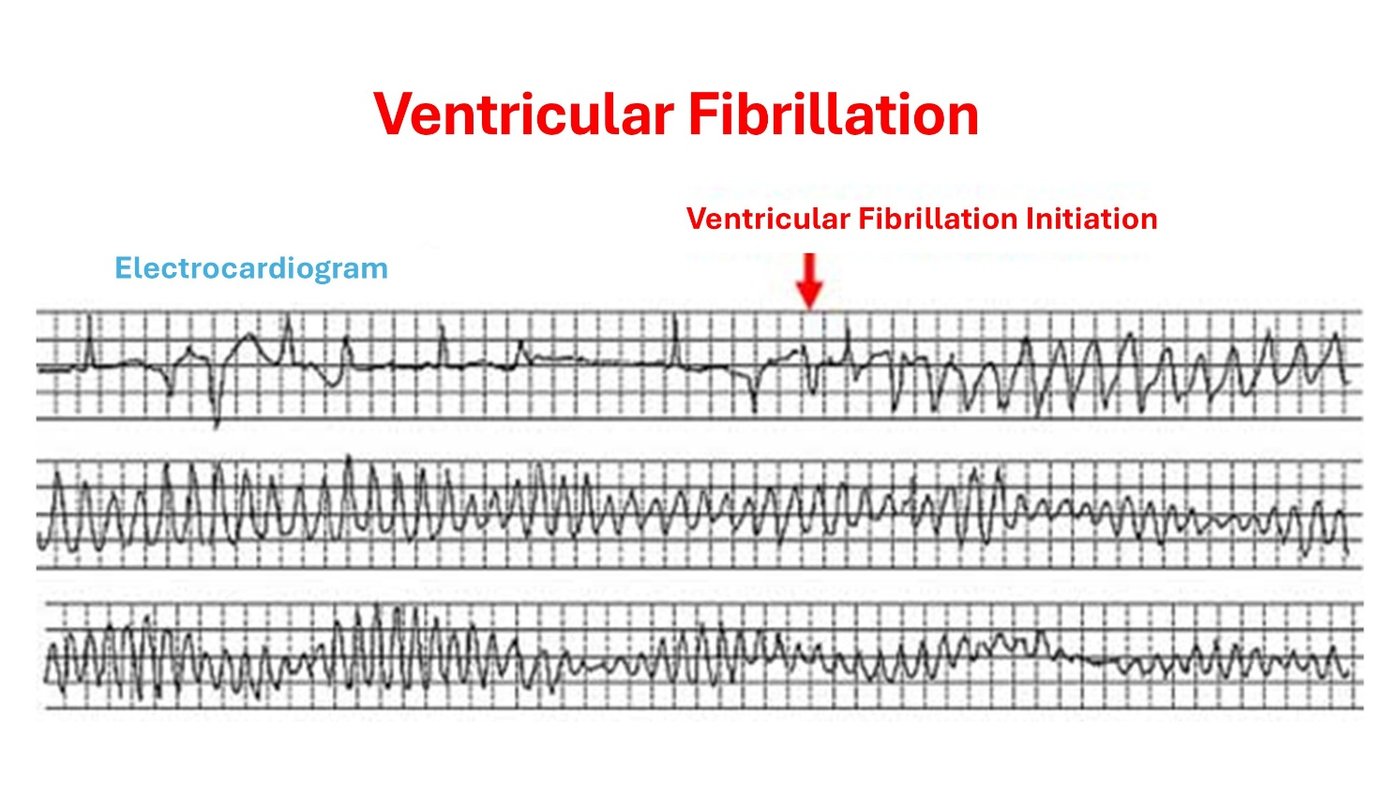
Cardiac conditions that can lead to sudden cardiac death
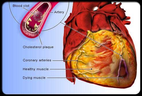
Acute Myocardial Infarction
In acute myocardial infarction, one of the arteries supplying the heart with oxygenated blood (a coronary artery) becomes abruptly occluded due to thrombosis triggered immediately after the rupture of an atheromatous plaque—a focal thickening located within the arterial lumen. Consequently, the region of the myocardium supplied by the specific artery is deprived of the oxygenated blood necessary for cardiomyocyte viability. One of the sequelae of this event, in certain cases, is myocardial electrical instability, which can culminate in life-threatening arrhythmias.
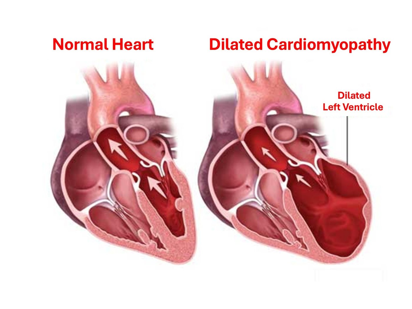
Cardiomyopathy
The term cardiomyopathy refers to structural or functional disorders of the heart muscle that impair its capacity to contract with the required force. This can result from various conditions, such as a prior myocardial infarction—referred to as ischemic cardiomyopathy—or ventricular dilation due to various non-coronary etiologies (e.g., viral infections, infectious agents, genetic causes), termed dilated or non-ischemic cardiomyopathy.
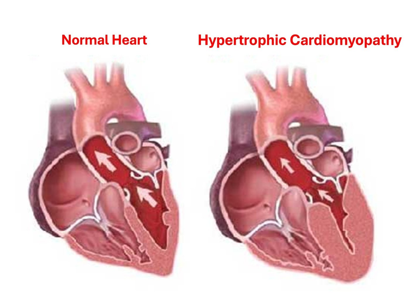
Hypertrophic Cardiomyopathy
This is a distinct form of cardiomyopathy characterized by hypertrophy (thickening) of the ventricular walls in the absence of external exacerbating factors (e.g., uncontrolled arterial hypertension, intense dynamic exercise). This thickening of the cardiac muscle is frequently responsible for disrupting the electrical activity of the heart, thereby precipitating severe arrhythmias.
Valvular Heart Diseases
In these cases, the cardiac valves—structures that ensure unidirectional blood flow—either exhibit insufficiency (permitting valvular regurgitation) or stenosis (obstructing normal blood flow). In severe cases where valvular dysfunction subjects the myocardium to excessive hemodynamic stress, the likelihood of severe arrhythmias increases.
Congenital Heart Diseases
Sudden death in children or adolescents is frequently attributable to congenital heart defects, which are structural anatomical anomalies of the heart present at birth. Furthermore, adults who underwent corrective surgery during childhood for certain congenital cardiac conditions may still carry a lifelong risk of sudden death.
Inherited Primary Arrhythmic Syndromes
In some individuals, the underlying abnormality precipitating the severe arrhythmia does not involve myocardial or valvular dysfunction but is purely electrical. This category comprises patients with inherited primary arrhythmic syndromes, occasionally referred to as channelopathies or electrical heart diseases. These are genetic disorders that induce electrical instability and severe cardiac arrhythmias. This group includes Brugada syndrome, Long QT syndrome, Short QT syndrome, early repolarization syndrome, and Catecholaminergic Polymorphic Ventricular Tachycardia (CPVT).
Beyond channelopathies, Wolff-Parkinson-White (WPW) syndrome is also classified among electrical cardiac disorders. In this syndrome, an accessory pathway—essentially an extra congenital electrical connection, or "wire"—connects the atria directly to the ventricles. This electrical bridge can facilitate the formation of various re-entrant arrhythmic circuits, the most severe of which involves the rapid conduction of atrial tachyarrhythmias (e.g., atrial fibrillation) from the atria to the ventricles. In such instances, the ventricles contract at an excessively high rate, ultimately degenerating into ventricular fibrillation and death. Fortunately, this probability remains low in patients with WPW syndrome; however, given its severity, it must never be overlooked during arrhythmic risk stratification.
Risk Factors
Given that 80% of sudden cardiac deaths occur in the setting of coronary artery disease, it follows that the risk factors for coronary artery disease concurrently serve as risk factors for sudden cardiac death.
- Smoking
- Family history of coronary artery disease
- Hypertension
- Elevated serum cholesterol levels
- Diabetes mellitus
- Obesity and a sedentary lifestyle
Other potential risk factors include:
- History of a prior cardiac arrest
- Low left ventricular ejection fraction (LVEF ≤ 40%)
- Family history of sudden cardiac death
- Use of illicit drugs and substances, such as cocaine, amphetamines, or other illegal agents
- Alcohol abuse
- Male sex
- Concurrently, a medical history positive for any of the aforementioned cardiac conditions and diseases etiologically linked to sudden cardiac death.
Sudden Death in Athletes
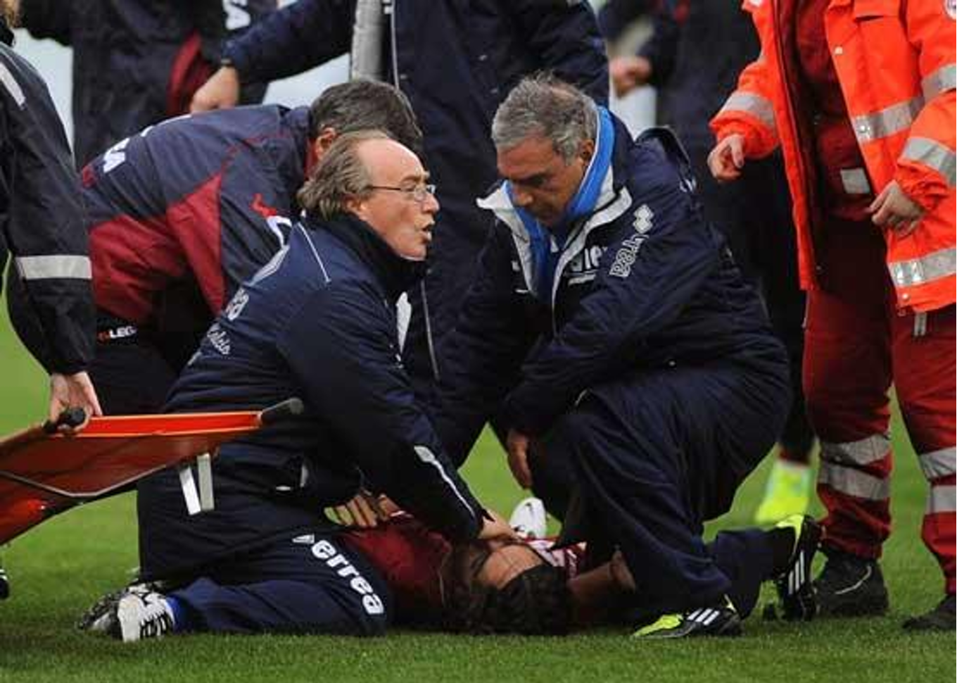 Whether the risk of sudden death is elevated in athletes remains unresolved by epidemiological data. Registries from Padua, Italy, reported an increased risk; however, these findings have not been replicated by other epidemiological studies across Europe and the United States.
Nevertheless, it is universally accepted that individuals diagnosed during pre-participation screening with a cardiac condition potentially capable of causing sudden death must discontinue competitive sports in an effort to mitigate this risk. This strategy, implemented over several years in Italy, appears to have reduced the incidence of sports-related sudden deaths from 3.6 per 100,000 individuals annually two decades ago, to 0.4 per 100,000 individuals annually today.
What should pre-participation screening entail? In our country, it has become standard practice in recent years for anyone engaging in sports—whether in childhood or later in life—to undergo a clinical cardiological evaluation, an electrocardiogram (ECG), and, if warranted (which practically applies to the majority), an echocardiogram.
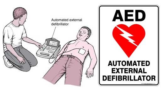
Another aspect demanding equal emphasis to pre-participation screening is the availability of automated external defibrillators (AEDs) at sports venues. Such a device, mounted in a prominent location analogous to a fire extinguisher, can save a life when the need arises. It is straightforward to use with minimal training, and, in our view, the physical presence of this device at sports complexes is potentially more valuable than that of a physician.
Diagnostic Evaluation
Patients who have been successfully resuscitated from a sudden cardiac death episode must undergo a meticulous diagnostic evaluation to uncover the underlying etiology and precisely stratify future risk, thereby enabling the implementation of measures to prevent recurrence.
Available diagnostic modalities include:
- The Electrocardiogram (ECG): Although a centenary diagnostic tool dating back over 100 years, it remains an essential instrument providing initial direction for the correct interpretation of the clinical event. An ECG can diagnose conditions such as acute infarction, QT interval prolongation, and electrolyte derangements.
- Blood Tests: These can identify electrolyte, metabolic, or endocrinological disorders, as well as confirm an acute myocardial infarction (via elevated serum cardiac biomarkers: cardiac enzymes, troponin) or an acute infection that further destabilizes the patient’s condition.
- Imaging Modalities: These may include chest radiography, echocardiography, cardiac magnetic resonance imaging (CMR), and myocardial perfusion scintigraphy (thallium scan). The primary objective of cardiac imaging is to delineate structural cardiac anomalies or detect myocardial ischemia.
- Other Diagnostic Procedures:
- Coronary Angiography: This remains the gold standard modality for identifying stenoses within the coronary arteries.
- Electrophysiology Study (EPS): An invasive procedure performed by introducing microscopic electrode catheters into specific locations within the cardiac chambers. It allows the assessment of the heart’s conduction system and evaluates the susceptibility of the myocardial substrate to sustain life-threatening tachyarrhythmias. It is rarely indicated in cases of documented episodes clearly caused by a severe ventricular tachyarrhythmia (e.g., ventricular tachycardia or fibrillation). Occasionally, it is performed to confirm a diagnosis or to stratify the risk of developing severe ventricular tachyarrhythmias.
Acute Management
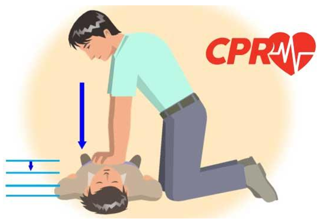
The vast majority of sudden cardiac death episodes occur in an out-of-hospital setting. In these scenarios, immediate cardiopulmonary resuscitation (CPR) is absolute imperative if there is to be any hope of survival. The objective of CPR is to sustain a baseline flow of oxygenated blood to vital organs, thereby establishing a critical physiological bridge until advanced life support becomes available.
If one witnesses such an event and is untrained in cardiopulmonary resuscitation, the primary action is to activate emergency medical services by calling 166. If the individual is apneic (not breathing), chest compressions must be initiated immediately, pushing hard and fast at a rate of 100 to 120 compressions per minute. This must continue until an automated external defibrillator (AED) is located or advanced medical assistance arrives.
How to Perform CPR
Should you encounter such an incident, the following logical steps must be executed sequentially:
- Determine whether the individual is conscious or unresponsive.
- If unresponsive, ask loudly: "Are you okay?"
- If the victim remains unresponsive and a second person is available, one should activate emergency services while the other immediately initiates CPR.
- If a lone rescuer has immediate access to a phone, they must call for help first and then initiate CPR, unless the victim’s cardiac arrest is secondary to asphyxia (e.g., drowning), in which case CPR should be performed for 1 minute prior to calling for assistance.
- If a lone rescuer encounters a pediatric victim, they must perform 2 minutes of CPR before calling for help or retrieving an AED.
- Initiate chest compressions at a rate of 100 to 120 compressions per minute.
- If untrained in CPR, continue continuous chest compressions (hands-only CPR) until professional help arrives.
- If trained in CPR, after 30 compressions, carefully perform a head-tilt/chin-lift maneuver to open the airway. Assess for spontaneous breathing for no more than 10 seconds. If breathing is absent, deliver 2 rescue breaths, ensuring visible chest rise. Pinch the victim’s nostrils closed, deliver the first rescue breath over one second, and observe the chest expansion. If the chest rises, deliver the second breath. If the chest fails to rise, readjust the head position before attempting the second rescue breath.
- If the victim is a child and fails to show signs of life after five cycles (approximately two minutes of CPR), retrieve and apply the AED. Deliver 1 shock if prompted by the device, and immediately resume chest compressions for 2 minutes before allowing the AED to analyze and potentially advise a 2nd shock. If untrained in AED operation, telephonic guidance can be requested from the emergency dispatcher by calling 166.
- Continue CPR until the individual shows signs of recovery and regains consciousness, or until professional help arrives.
Long-Term Management
Following successful resuscitation from such an episode, a exhaustive investigation into the precipitating cause must be conducted. Once the etiology is identified, comprehensive measures must be implemented to shield the patient from the recurrent risk of sudden death. Generally, therapeutic strategies must target the optimal management of the underlying cardiac disease from which the averted sudden death event arose.
Regardless of the specific cardiac pathology, the implantable cardioverter-defibrillator (ICD) plays a paramount role in therapy. This is a miniaturized, battery-powered device implanted via a surgical incision below the clavicle into a subcutaneous pocket. The device is connected to 1 to 3 leads (depending on the configuration) which are threaded transvenously into the cardiac chambers. The ICD resembles a standard pacemaker and can function as such by emitting pacing stimuli to induce myocardial contraction. Concurrently, it continuously senses and analyzes the cardiac rhythm, delivering immediate therapy to terminate life-threatening arrhythmias.
A modern alternative is the subcutaneous defibrillator (S-ICD). In contrast to conventional transvenous systems, this device and its lead are positioned entirely subcutaneously over the thorax, avoiding any lead insertion into the venous system or the heart itself. This significantly minimizes the risk of systemic intravascular infections or lead fractures. However, the S-ICD is utilized exclusively for terminating dangerous tachyarrhythmias via electrical shock (defibrillation) and lacks the capacity to provide permanent bradycardia pacing.
Other therapeutic measures that may be implemented include:
- Pharmacotherapy: Beta-blockers, angiotensin-converting enzyme (ACE) inhibitors, mineralocorticoid receptor antagonists (spironolactone, eplerenone), or amiodarone may be prescribed as indicated.
- Percutaneous coronary intervention (PCI) and stent implantation in the presence of coronary artery stenosis or acute myocardial infarction.
- Coronary artery bypass grafting (CABG).
- Surgical valve repair or replacement if valvular dysfunction is the primary causative factor.