
Arrhythmogenic Right Ventricular Cardiomyopathy (ARVC) is a genetic disorder of the cardiac muscle. This means it can be inherited and is caused by a mutation in one or more genes. The disease primarily affects the right ventricle of the heart—the chamber that pumps blood to the lungs. However, it very frequently involves the left ventricle to a lesser extent, which is perhaps the most vital of the four cardiac chambers, responsible for pumping oxygenated blood through the arteries to the rest of the body.
A Greek cardiologist from Naxos, Nikos Protonotarios, made a significant contribution to the discovery of the disease. In 1986, he investigated a series of patients presenting with right ventricular cardiomyopathy, arrhythmias, a characteristic hyperkeratosis of the palms and soles, and unusually curly hair. This specific variant of the disease was named "Naxos disease". It was later discovered to be caused by a mutation in plakoglobin, a protein that links cardiac cells together.
What is the cause of the disease?
Arrhythmogenic Right Ventricular Cardiomyopathy is caused by defective proteins that bind cardiac cells together. These defective proteins result from mutations present in specific genes, which are transmitted hereditarily.
Normally, cardiac cells are linked together by strong protein bonds (Figure 1A). If this connection is defective, cellular cohesion is disrupted, particularly under conditions of stress (Figure 1B). In its attempt to heal this injury, the body deposits fibrous tissue and fat in the intervening areas (Figure 1C). This disruption in cellular cohesion impairs the transmission of the electrical impulse—meaning the unobstructed electrical communication between cells—and is responsible for generating ventricular arrhythmias, which can sometimes prove fatal.
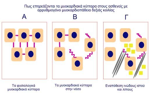
Figure 1
- A: Normal myocardial cells
- B: Myocardial cells in disease state
- Γ: Deposition of fibrous tissue and fat
How is the disease inherited?
Our body contains trillions of cells. Each cell has its own nucleus, which contains all the information that makes each of us unique. This information is encoded in our genes (Figure 2).
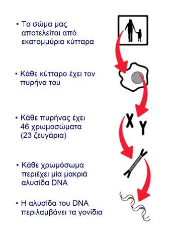
Figure 2
- The body consists of millions of cells.
- Each cell contains its own nucleus.
- Each nucleus contains 46 chromosomes (23 pairs).
- Each chromosome contains a long strand of DNA.
- The DNA strand includes the genes.
Figure 2
We inherit one set of chromosomes, and therefore one set of genes, from each parent to form pairs. If a certain gene carries an error—that is, a mutation—it can produce a defective protein. This protein performs a defective function in the organism, leading to a disease such as Arrhythmogenic Right Ventricular Cardiomyopathy. This mutation in the specific gene is transmitted to subsequent generations. Whether a mutation will manifest as clinical disease depends on many, often unknown, factors.
One of the common inheritance patterns in Arrhythmogenic Right Ventricular Cardiomyopathy is autosomal dominant. Each child has a 50% probability of inheriting the mutation from an affected parent. However, not everyone who carries the mutation will necessarily develop the disease. This phenomenon is known as incomplete penetrance (see Figure 3). Consequently, the disease appears to skip certain generations and manifest in subsequent ones.
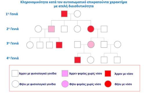
Figure 3. Pedigree chart. Autosomal dominant inheritance pattern with incomplete penetrance.
- 1st Generation / 2nd Generation / 3rd Generation / 4th Generation
- Male with normal genes / Female with normal genes
- Male unaffected carrier / Female unaffected carrier
- Male with clinical disease / Female with clinical disease
Another mode of inheritance encountered in Arrhythmogenic Right Ventricular Cardiomyopathy is autosomal recessive. This means that for an individual to develop the clinical disease, they must inherit two pathological genes. Furthermore, if an individual is a carrier, they do not suffer from the disease but can transmit it (see Figure 4).
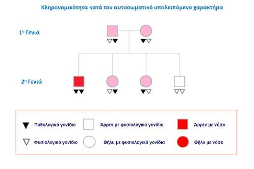
Figure 4. Pedigree chart. Autosomal recessive inheritance pattern.
- 1st Generation / 2nd Generation
- Pathological gene / Normal gene
- Male with normal genes / Female with normal genes
- Male with clinical disease / Female with clinical disease
Symptoms
Patients may frequently be asymptomatic or present with minimal symptoms. At other times, they may exhibit a range of symptoms:
- Palpitations: Primarily caused by ventricular premature complexes or brief episodes of tachycardia.
- Lightheadedness or Syncope: This symptom is also typically a consequence of arrhythmias.
- Edema (primarily of the lower extremities): This occurs when the right ventricle exhibits such structural dysfunction that it cannot effectively pump blood, leading to fluid retention.
- Dyspnea (exertional dyspnea): This can be caused by right ventricular dysfunction, arrhythmias, as well as left ventricular dysfunction.
Arrhythmias
Due to the structural abnormalities described above, the heart’s electrical impulse can be disrupted, leading to arrhythmias:
- Ventricular Premature Complexes (VPCs): This is the most common arrhythmia in Arrhythmogenic Right Ventricular Cardiomyopathy. These are contractions that interrupt the normal rhythm, originating from the ventricles (primarily the right ventricle). They do not follow the physiological pathway of the cardiac electrical impulse, which normally travels from the atria to the ventricles.
- Ventricular Tachycardia (VT): A sustained rhythm originating from the heart ventricles. It may be short in duration (up to 30 seconds), termed non-sustained, or persist longer, in which case it is termed sustained.
- Ventricular Fibrillation (VF): A potentially fatal arrhythmia if not treated immediately with defibrillation (an electrical shock that restores normal heart rhythm).
- Atrial Fibrillation (AF): An arrhythmia of the atria that causes the heart to beat irregularly and rapidly. It is usually not immediately life-threatening, but it can cause symptoms and predispose patients to thrombus formation and embolic/stroke events.
Diagnosis
The diagnosis of Arrhythmogenic Right Ventricular Cardiomyopathy is based on medical history, family history, physical examination, and diagnostic tests such as: electrocardiogram (ECG), signal-averaged ECG, Holter monitoring, echocardiography, and cardiac magnetic resonance imaging (MRI) or computed tomography (CT). Additionally, an electrophysiology study, coronary angiography (to exclude coronary artery disease), or, more rarely, histological evaluation via endomyocardial biopsy may be required.
Cardiac MRI is perhaps the imaging modality with the highest accuracy for visualizing suspicious and pathological areas in the myocardium of both the right and left ventricles. Unfortunately, it cannot be performed on patients who already have an implanted defibrillator (although today we implant MRI-compatible defibrillators); however, diagnosis is typically established prior to defibrillator implantation.
The key criteria to be evaluated that increase diagnostic certainty include:
- Evident myocardial structural abnormalities of the right ventricle
- Abnormal electrocardiogram
- Presence of arrhythmias highly indicative of the disease
- Family history of the condition
Treatment
The management of Arrhythmogenic Right Ventricular Cardiomyopathy is directed toward preventing the risk of life-threatening arrhythmias, improving symptoms arising from structural disorders, and halting disease progression. Therapeutic interventions may include:
- Lifestyle Modifications
Patients must avoid strenuous exertion and competitive sports. Apart from the risk of triggering arrhythmias, intense exercise has been implicated in the adverse progression of the disease. - Pharmacotherapy
Treatment to manage right or left ventricular heart failure (beta-blockers, angiotensin-converting enzyme inhibitors, diuretics). Antiarrhythmic therapy, such as amiodarone, to suppress arrhythmias. - Defibrillator Implantation
An implantable cardioverter-defibrillator (ICD) is a miniature device placed via an incision in the skin below the clavicle. It is connected to one or two leads guided through the subclavian vein into the interior of the heart. The ICD continuously monitors the cardiac rhythm, and if it detects a severe arrhythmia, it activates to terminate it via antitachycardia pacing or electrical discharge (shock) [0.1.6–0.1.7].
ICD implantation is indicated when severe arrhythmias have already occurred (ventricular tachycardia, ventricular fibrillation) or prophylactically when the patient exhibits high-risk features for the onset of such dangerous arrhythmias, such as severe structural right ventricular impairment, left ventricular dysfunction, or prior syncope.
- Catheter Ablation
Ablation therapy is considered when a patient experiences recurrent episodes of ventricular tachycardia and potentially frequent appropriate shocks from the ICD. For ablation, three-dimensional cardiac mapping systems are utilized, allowing detailed visualization of the electrical voltage at every point of the heart, thereby identifying pathological areas (Figure 5). Utilizing these technological advances, precise interventions can be performed in the specific regions responsible for the genesis and maintenance of arrhythmias. - Heart Transplantation
This option is reserved for patients with end-stage heart failure or refractory, incessant arrhythmias that cannot be suppressed by pharmacotherapy, the ICD, or catheter ablation performed at experienced centers.
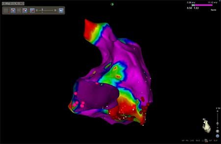
Figure 5. Image from a clinical case in our electrophysiology laboratory. The right ventricle is visualized using a three-dimensional mapping system displaying the electrical voltage at each site. Regions in red represent low-voltage areas where disease is present.