
Definition
Hypertrophic cardiomyopathy (HCM) is a myocardial disease characterized by a pathological thickening (hypertrophy) of the ventricular walls. It predominantly possesses a genetic etiology, arising from pathogenic gene variants transmitted across generations via hereditary patterns.
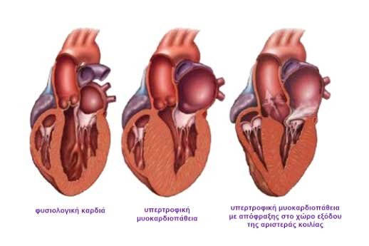
Figure 1
In a physiologically normal heart, cardiomyocytes are arranged in an organized, parallel alignment. Conversely, in hypertrophic cardiomyopathy, they exhibit a highly irregular, disorganized structural pattern, a phenomenon medically defined as "myocardial disarray". Consequently, the cardiac walls—primarily those of the left ventricle—become thick and non-compliant (stiff), which can significantly compromise mechanical cardiac function.

In certain clinical presentations, the localized myocardial thickening induces a narrowing within the left ventricular outflow tract (LVOT)—the specific anatomical pathway through which blood is ejected into the aorta and the systemic circulation. These cases are classified as "obstructive hypertrophic cardiomyopathy".
Beyond HCM, which stands as an inherited primary genetic disorder, secondary myocardial hypertrophy can be induced by other clinical etiologies. These include chronic, uncontrolled arterial hypertension, aortic valve stenosis, or long-term, high-intensity resistance training in strength sports such as powerlifting and wrestling.
As a primary genetic disorder caused by mutations affecting the sarcomere, HCM typically manifests phenotypically during adolescence. Clinical diagnosis is established through patient symptomatology, the detection of a cardiac murmur during auscultation, or abnormal, suspicious findings on a resting electrocardiogram (ECG).
Hereditary Transmission
The disease is typically inherited in an autosomal dominant pattern. This implies that an offspring of an affected individual has a 50% probability of inheriting the pathogenic variant. This hereditary transmission can be mapped and evaluated by constructing a comprehensive medical pedigree chart. Family members who do not inherit the genetic mutation cannot transmit the disease to their descendants. Furthermore, individuals who do inherit the pathogenic variant exhibit variable expressivity, meaning they will not necessarily demonstrate the same degree of clinical severity or symptomatology.
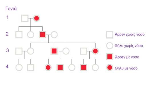
Figure 3: Medical pedigree chart depicting the autosomal dominant inheritance pattern of hypertrophic cardiomyopathy.
Symptomatology
A significant cohort of patients diagnosed with hypertrophic cardiomyopathy remain entirely asymptomatic and maintain a normal life expectancy. Conversely, other patients develop clinical symptoms that may progressively worsen alongside disease evolution and myocardial remodeling.
Symptoms, which can manifest at any stage of life, include:
- Chest pain or angina-like chest heaviness: Predominantly triggered by physical exertion, though it may also occur at rest, particularly postprandially following large meals.
- Dyspnea: Shortness of breath, characteristically exacerbated during physical exertion.
- Asthenia and fatigue: Generalized weakness and reduced exercise tolerance.
- Syncope or presyncope: Fainting episodes that may arise from severe cardiac arrhythmias, abnormal vascular responses (hemodynamic instability) during exertion, or other cryptogenic etiologies.
- Palpitations: Subjective awareness of an irregular heartbeat, frequently originating from atrial fibrillation or ventricular arrhythmias.
- Sudden Cardiac Death (SCD): Precipitated by malignant ventricular arrhythmias; fortunately, this occurs in a small percentage of the patient population.
Diagnostic Evaluation
The diagnosis of hypertrophic cardiomyopathy is established based on a thorough personal and family medical history, physical examination, electrocardiography (ECG), and transthoracic echocardiography. Supplementary diagnostic modalities may include specialized blood panels, exercise stress testing, cardiac magnetic resonance imaging (CMR), cardiac computed tomography (CT), and coronary angiography.
Hypertrophic Cardiomyopathy and Arrhythmogenesis
Myocardial hypertrophy, the disarray of cardiomyocytes, and interstitial fibrosis are primary pathophysiological substrates that render HCM patients highly susceptible to both atrial and ventricular tachyarrhythmias.
The most severe clinical consequence of these arrhythmias is Sudden Cardiac Death (SCD), primarily triggered by malignant ventricular tachyarrhythmias such as ventricular fibrillation. HCM represents the leading cause of SCD in young individuals and competitive athletes under the age of 35.
Specifically, the arrhythmic profile associated with hypertrophic cardiomyopathy includes:
- Atrial Fibrillation (AF): A supraventricular tachyarrhythmia characterized by rapid, disorganized atrial activation, effectively compromising atrial mechanical function. AF results in a rapid, irregular ventricular response that further impairs diastolic filling and exacerbates clinical symptoms. Furthermore, AF significantly elevates the risk of thromboembolic events, specifically ischemic stroke. Consequently, patients with HCM and documented AF must immediately initiate oral anticoagulation therapy. Notably, standard clinical risk stratification models, such as the CHA₂DS₂-VASc score, are not applicable for risk assessment in this patient population.
- Non-Sustained Ventricular Tachycardia (NSVT): Defined as a rapid ventricular rhythm (>100 beats per minute) lasting less than 30 seconds. While usually asymptomatic, the presence of NSVT on ambulatory Holter monitoring is a recognized clinical risk factor—a warning sign—for sustained, life-threatening ventricular arrhythmias that can precipitate SCD.
- Premature Ventricular Complexes (PVCs): Isolated, ectopic beats originating from the ventricles. They are highly prevalent in HCM patients and can stem from diverse etiologies. Their detection necessitates rigorous, extended ambulatory ECG (Holter) monitoring to screen for underlying NSVT episodes.
- Sustained Ventricular Tachycardia: Monomorphic sustained ventricular tachycardia occurs infrequently in HCM, typically presenting in advanced stages of the disease as a result of extensive myocardial scarring and localized fibrosis.
- Ventricular Fibrillation and Sudden Death: A catastrophic, life-threatening ventricular arrhythmia that causes hemodynamic collapse and results in fatal cardiac arrest within minutes unless immediate cardiopulmonary resuscitation and defibrillation are performed. It remains the primary clinical concern in HCM management. It can occur at any stage of the disease, even as the initial manifestation before a formal diagnosis is established or symptoms appear. Fortunately, it affects a small but statistically significant percentage of patients. Longitudinal cohort studies have identified specific clinical markers for SCD risk. Contemporary risk stratification allows clinicians to estimate this risk and, when indicated, proceed with the implantation of an Implantable Cardioverter-Defibrillator (ICD), which represents the only definitive preventive therapeutic intervention.
Risk factors for SCD include extreme left ventricular wall thickness, documented episodes of NSVT, a history of unexplained syncope, a family history of premature SCD in a first-degree relative, left atrial enlargement, and a high left ventricular outflow tract (LVOT) gradient.
The European Society of Cardiology (ESC) provides an online risk calculator (doc2do.com) to estimate the 5-year risk of sudden cardiac death, guiding clinical decision-making regarding prophylactic ICD implantation.
Therapeutic Management
The therapeutic strategy for hypertrophic cardiomyopathy focuses on symptom palliation, slowing disease progression, and preventing adverse cardiovascular events, specifically lethal arrhythmias and sudden death.
Targeted interventions are tailored to the clinical stage and disease severity, encompassing:
- Lifestyle Modifications: Patients diagnosed with HCM must strictly abstain from competitive sports. Depending on disease severity, low-intensity aerobic exercise may be permitted. Adequate hydration is highly recommended, except in advanced stages characterized by severe congestive heart failure.
- Pharmacotherapy: Medical therapy aims to optimize diastolic filling by reducing heart rate and enhancing myocardial relaxation. First-line agents include beta-blockers (e.g., metoprolol, propranolol, atenolol) and non-dihydropyridine calcium channel blockers (e.g., verapamil, diltiazem). For rhythm control in patients with atrial or ventricular arrhythmias, amiodarone may be indicated. In the presence of atrial fibrillation, oral anticoagulants must be administered, including vitamin K antagonists (e.g., acenocoumarol) or Direct Oral Anticoagulants (DOACs) such as dabigatran, rivaroxaban, or apixaban.
- Implantable Cardioverter-Defibrillator (ICD) Implantation: ICD therapy serves as the most effective strategy for the secondary prevention of SCD in patients who have survived a cardiac arrest or sustained ventricular tachycardia. Prophylactic (primary prevention) ICD implantation is indicated in asymptomatic or minimally symptomatic patients presenting with high-risk clinical features. According to ESC guidelines, patients with an estimated 5-year SCD risk score of ≥6% are candidates for ICD therapy.
- Surgical Septal Myectomy: An open-heart surgical procedure involving the resection of a portion of the hypertrophied interventricular septum that causes LVOT obstruction. This specialized intervention is performed at dedicated centers of excellence and is reserved for patients with refractory symptoms despite maximal tolerated medical therapy.
- Alcohol Septal Ablation: A percutaneous catheter-based intervention involving the targeted infusion of absolute alcohol into a selective septal perforator branch of the coronary arteries. This induces a controlled myocardial infarction, reducing septal mass and alleviating LVOT obstruction. Potential complications include the development of a high-grade atrioventricular (AV) block, which may necessitate permanent pacemaker implantation.
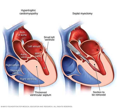
Figure 4: Comparative medical illustration demonstrating hypertrophic cardiomyopathy versus a surgical septal myectomy.