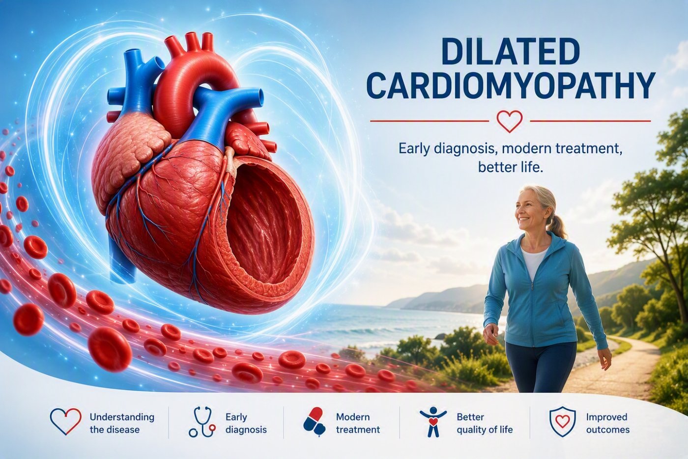
Definition
Dilated cardiomyopathy (DCM) is a severe myocardial disease characterized by the dilation (enlargement) of the cardiac chambers, primarily affecting the left ventricle. As the ventricular walls stretch, they become thin and weakened, severely compromising the heart’s contractile capacity and its ability to effectively pump blood throughout the systemic circulation. Unlike ischemic cardiomyopathy, this myocardial injury is not secondary to coronary artery disease (stenoses). Instead, it is precipitated by a diverse array of etiologies, most notably genetic mutations, family history, viral infections (myocarditis), chronic alcohol abuse, cardiotoxic agents (such as specific chemotherapeutic regimens), or autoimmune disorders.
Pathophysiological Impact on Cardiac Function
Triggered by primary myocardial cell injury or sustained elevated wall stress, the heart initiates a maladaptive process known as ventricular remodeling. While the heart initially attempts to compensate for the diminished inotropic capacity, this compensatory mechanism ultimately establishes a deleterious pathway that exacerbates cardiac dysfunction:
- Dilation and Thinning: The left ventricular wall undergoes progressive eccentric hypertrophy and dilation (stretching) to accommodate increased end-diastolic volumes, attempting to maintain systemic cardiac output.
- Spherical Structural Shift: As dilation advances, the left ventricle loses its physiological ellipsoid geometry and transitions into a more spherical shape, severely impairing its mechanical and hemodynamic efficiency.
- Deleterious Feedback Loop: Progressive dilation escalates ventricular wall tension and increases myocardial workload. This leads to a relentless advancement of heart failure and promotes diffuse myocardial fibrosis (scarring) within the cardiac tissue.
Modern pharmacological strategies—including beta-blockers, ACE inhibitors/ARNI, and SGLT2 inhibitors—specifically target the inhibition or potential reversal of this maladaptive remodeling process (reverse remodeling), facilitating chamber reduction and restoring partial myocardial functionality.
Clinical Presentation
While many patients remain asymptomatic during the early clinical stages, the progression of the disease manifests through classic symptoms of congestive heart failure:
- Dyspnea: Exertional shortness of breath, which may progress to orthopnea (difficulty breathing when supine) or dyspnea at rest.
- Fatigue and Exercise Intolerance: Marked generalized weakness, lethargy, and a significantly reduced functional capacity.
- Peripheral and Systemic Edema: Swelling in the lower extremities (ankles and feet) or abdominal ascites.
- Syncope and Vertigo: Transient loss of consciousness (fainting) or dizziness.
- Palpitations: Sensations of skipped beats, fluttering, or atypical chest discomfort secondary to underlying arrhythmias.
Diagnostic Evaluation
A definitive diagnosis is established based on a thorough medical history, physical examination, and a structured multimodality diagnostic workup:
- Electrocardiogram (ECG): Reveals evidence of chamber hypertrophy, intraventricular conduction delays such as Left Bundle Branch Block (LBBB), or complex arrhythmias.
- Echocardiography (Transthoracic Triplex): The primary diagnostic modality used to confirm DCM by directly assessing chamber dilation, wall thickness, and left ventricular ejection fraction (LVEF).
- Cardiovascular Magnetic Resonance (CMR): A critical imaging tool for evaluating DCM. CMR identifies the presence and precise distribution of myocardial fibrosis via late gadolinium enhancement (LGE), assisting in the differentiation between ischemic and non-ischemic etiologies.
- Coronary Angiography: Indicated to conclusively rule out obstructive coronary artery disease as the primary cause of left ventricular dysfunction.
- Genetic Testing: Recommended, particularly in suspected familial or hereditary forms, to identify specific pathogenic mutations underlying the disease.
Dilated Cardiomyopathy and Arrhythmogenesis
Structural alterations of the myocardium, chamber dilation, and diffuse interstitial fibrosis disrupt regular electrical conduction. This generates a state of electrical instability and provides the necessary substrate for dangerous re-entrant arrhythmias:
- Ventricular Tachycardia (VT) and Ventricular Fibrillation (VF): These represent life-threatening malignant ventricular arrhythmias and serve as the leading cause of Sudden Cardiac Death (SCD) in this patient cohort. The risk is heavily compounded by the extent of myocardial fibrosis identified on CMR.
- Atrial Fibrillation (AF): Frequently precipitated by atrial dilation and elevated filling pressures. AF severely compromises the already failing heart, accelerates heart failure exacerbations, and dramatically increases thromboembolic stroke risks, necessitating mandatory oral anticoagulation therapy.
- Premature Ventricular Contractions (PVCs): Extremely common ectopy requiring continuous ambulatory ECG monitoring (Holter monitor) to screen for non-sustained ventricular tachycardia or high arrhythmic burdens.
- Conduction System Disease: Myocardial degeneration often involves the cardiac conduction pathways, leading to abnormalities such as Left Bundle Branch Block (LBBB). LBBB induces mechanical dyssynchrony, further undermining ventricular performance.
Therapeutic Management
Therapeutic interventions are aimed at halting disease progression, alleviating clinical symptoms, optimizing cardiac output, and mitigating the risk of sudden cardiac death:
- Lifestyle Modifications: Absolute cessation of alcohol consumption (as it can be an immediate etiological factor or major disease driver), smoking cessation, strict blood pressure control, dietary modifications (essential restriction of sodium and fluid intake), and tailored, medically supervised exercise programs.
- Pharmacological Therapy: Medical management represents the cornerstone of DCM treatment. Pharmacotherapy directly targets pathophysiological pathways to protect the myocardium, prevent malignant arrhythmias, and substantially reduce hospitalization rates. Management centers on the guideline-directed medical therapy (GDMT) known as the "Golden Quadruplet" (The Big Four). These agents must be used in combination and uptitrated progressively to maximum tolerated target doses to facilitate reverse remodeling:
- Angiotensin Receptor-Neprilysin Inhibitors (ARNI): Sacubitril/Valsartan (Entresto). Replaces standard ACE inhibitors to provide superior neurohormonal blockade and cardiovascular protection.
- Beta-blockers (β-blockers): Reduce myocardial workload, lower heart rate, and protect against lethal arrhythmias. Extensively used agents include metoprolol succinate, carvedilol, bisoprolol, and nebivolol.
- Mineralocorticoid Receptor Antagonists (MRA): Spironolactone or eplerenone. These agents reduce fluid retention and suppress profibrotic pathways to minimize scar tissue formation.
- SGLT2 Inhibitors (Gliflozins): Dapagliflozin or empagliflozin. These provide profound clinical benefits, significantly lowering heart failure hospitalizations and cardiovascular mortality.
- Adjunctive Medical Therapy: The foundational quadruplet must be complemented by specialized medications critical for patient stabilization:
- Diuretics (e.g., Furosemide): The cornerstone of daily fluid volume management. They are crucial for immediate decongestion of pulmonary and systemic volume overload, rapidly relieving dyspnea, preventing emergency admissions for acute pulmonary edema, and enabling the safe initiation and uptitration of GDMT.
- Anticoagulants (e.g., Apixaban, Rivaroxaban, Dabigatran): Mandated in patients with concomitant atrial fibrillation to provide robust protection against ischemic stroke.
- Antiarrhythmic Drugs (e.g., Amiodarone): Employed to manage high arrhythmic burdens and suppress recurrent ventricular episodes.