
Definition
Atrial tachycardia is a type of supraventricular tachycardia that originates within, and has its focus in, one of the two atria of the heart. This arrhythmogenic focus typically exhibits an impulse firing rate exceeding 100 beats per minute, causing the heart to contract accordingly. This high frequency suppresses the heart’s physiological pacemaker, namely the sinus node, which becomes overridden by the rapid rate of the atrial tachycardia and goes silent. For the normal sinus rhythm to re-emerge, the atrial tachycardia must terminate.
Mechanisms
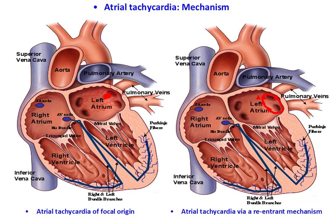
Atrial tachycardia can be generated via two distinct mechanisms:
- Focal Origin: The tachycardia originates from a small, localized region within the right or left atrium that fires at a high frequency.
- Re-entrant Mechanism: The tachycardia is sustained by the continuous and rapid circus-movement propagation of an electrical impulse around a discrete anatomical or functional circuit. This rapid activation subsequently drives the remainder of the atrial myocardium (see video).
Symptoms
The symptomatic presentation of atrial tachycardia mirrors that of other supraventricular tachycardias and includes:
- Palpitations
- Dizziness / Lightheadedness
- Asthenia / Weakness
- Dyspnea
- Reduced exercise tolerance
- Chest discomfort
Etiology
In many instances, this condition occurs in individuals with a structurally normal heart who present with no other demonstrable cardiac pathology, even following an extensive diagnostic workup. In contrast to atrial fibrillation, atrial tachycardia rarely manifests during the acute phase of cardiac disorders or during the acute exacerbation of pre-existing cardiovascular diseases.
The etiological factors underlying atrial tachycardia may encompass the following conditions:
- Atrial tachycardia may result from localized necrosis and subsequent scar formation within the atrial myocardium, stemming from a historical viral infection or myocarditis. This myocardial scar provides the substrate necessary for the development of re-entrant circuits.
- It may occur in patients post-cardiac surgery, arising as a sequela of surgical incisions and interventions performed within the atria.
- In patients with atrial fibrillation who are treated with antiarrhythmic drugs (such as amiodarone, sotalol, propafenone, or flecainide), the atrial fibrillation can occasionally organize and transform into atrial tachycardia. If a concomitant AV-nodal blocking agent has not been prescribed to slow atrioventricular conduction, this rapid atrial activation can be conducted down to the ventricles via the AV node, culminating in a tachycardia with a ventricular rate substantially higher than that observed during the initial atrial fibrillation.
- Atrial tachycardia can also develop in patients who have undergone catheter ablation for atrial fibrillation. This is typically a persistent form of tachycardia that requires management via a repeat ablation procedure.
Diagnosis
The diagnosis of atrial tachycardia is established via a standard surface electrocardiogram (ECG) or ambulatory Holter monitoring. On a surface ECG, atrial tachycardia can occasionally mimic other supraventricular tachycardias, such as atrioventricular nodal re-entrant tachycardia (AVNRT) or certain macro-reentrant tachycardias involving an accessory pathway. In these scenarios, subtle electrocardiographic features, identified through clinical experience, can provide the differential diagnosis.
Diagnostic challenges frequently arise because episodes are often paroxysmal and short-lived, resolving before a standard ECG can be recorded. An ECG performed after the episode has terminated typically demonstrates normal sinus rhythm. Furthermore, structural and functional evaluations may also yield completely normal results; thus, the clinical history often provides the primary diagnostic suspicion. More frequent electrocardiographic screening, the immediate recording of an ECG at the onset of symptoms, and, in rarer cases, invasive diagnostic procedures such as an electrophysiology study (EPS) can definitively confirm the diagnosis. An EPS is the gold-standard investigation to precisely delineate the arrhythmia mechanism and map the arrhythmogenic focus. Whenever an EPS is scheduled, the electrophysiology laboratory must concurrently be prepared to deliver definitive therapy via catheter ablation.
Prognosis
Atrial tachycardia is a rhythm disorder that is generally not life-threatening. However, patients may experience severe, highly distressing symptoms that provoke significant anxiety. In such cases, clinical management is strongly indicated. Less frequently, a chronic, rapid ventricular rate can impair myocardial contractility, culminating in a form of heart failure termed "tachycardia-induced cardiomyopathy" (or tachycardiomyopathy). In these clinical scenarios, aggressive and decisive intervention is mandatory.
Management
Therapeutic strategy in patients with atrial tachycardia is dictated by the frequency of episodes, the presence and severity of symptoms, and the overall impact of the tachycardia on the patient’s cardiovascular status.
Therapeutic interventions aim to achieve:
- Acute termination (cardioversion) of symptomatic episodes
- Rate control (normalization of the ventricular response)
- Long-term maintenance of normal sinus rhythm
These clinical endpoints can be achieved via:
- Pharmacological therapy
- Catheter ablation
Pharmacological agents administered for ventricular rate control—which decrease the ventricular rate while the underlying atrial tachycardia persists—include beta-blockers, non-dihydropyridine calcium channel blockers, and, less frequently, digoxin.
Antiarrhythmic drugs prescribed to prevent recurrent episodes or to acutely terminate an active tachycardia episode include Class I and Class III agents, such as amiodarone, sotalol, propafenone, and flecainide. The specific agent that will prove clinically effective cannot be predicted a priori through initial testing or diagnostic procedures. Consequently, multiple sequential therapeutic trials may be required to identify the optimal drug regimen capable of effectively suppressing the arrhythmia.
Patients can achieve significant symptom relief using either of the aforementioned pharmacological approaches.
However, antiarrhythmic medications are frequently ineffective, fail to suppress symptoms to a satisfactory degree, or cause drug intolerance and adverse effects. In these instances, catheter ablation offers a radical, definitive, and curative therapeutic solution. Catheter ablation is typically indicated following the failure of medical therapy, upon the development of drug-induced side effects, or when the patient prefers to avoid long-term, chronic pharmacological therapy.