
Introduction
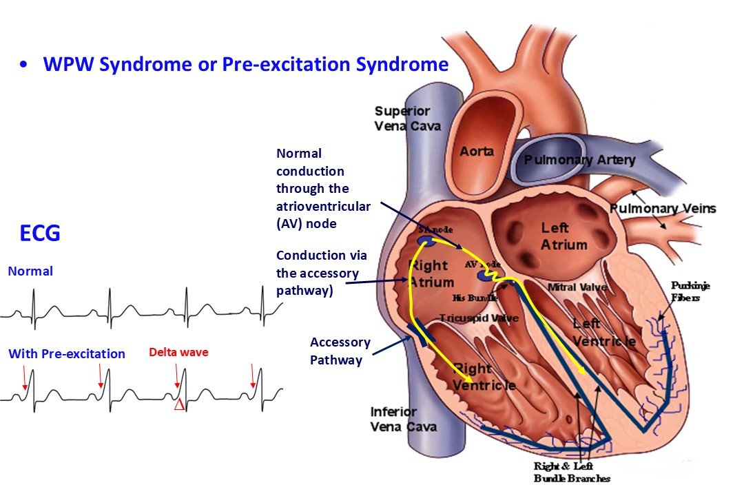
The underlying cause of these tachycardias is the presence of an accessory pathway. Under normal physiological conditions, the atrioventricular (AV) node and the bundle of His constitute the sole electrical conduit between the atria and the ventricles (see "The Electrical System of the Heart"). However, certain individuals—who otherwise typically present with a structurally normal heart—possess an anomalous electrical connection between the atria and the ventricles, resembling an extra micro-wire.
This anomalous atrioventricular connection (referred to as an accessory or ectopic pathway) typically represents an embryological remnant from the period of fetal cardiac development. While it may spontaneously involute during childhood or adolescence, it can otherwise persist throughout adulthood if left untreated.
The presence of manifest accessory pathways can be readily diagnosed on a standard 12-lead electrocardiogram (ECG), with the exception of concealed pathways. When this electrical substrate is associated with clinical symptomatology, the condition is termed Wolff-Parkinson-White (WPW) syndrome, named after the cardiologists Louis Wolff (1898–1972), John Parkinson (1885–1976), and Paul Dudley White (1886–1973), who comprehensively described this clinical entity in 1930.
Arrhythmia Mechanisms
Unlike the AV node, this accessory pathway does not possess decremental conduction properties (the ability to delay electrical impulses traveling from the atria to the ventricles); instead, it exhibits rapid, high-velocity conduction.
Consequently, during normal sinus rhythm, the ventricles undergo pre-excitation, as the cardiac impulse reaches them more rapidly via the accessory pathway than through the physiological conduction system (the AV node). This is why WPW syndrome is frequently designated as a pre-excitation syndrome.
On the ECG, ventricular pre-excitation manifests as a slurred upstroke at the onset of the otherwise steep QRS complex deflection (see "Electrocardiogram"), which corresponds to ventricular depolarization (Figure 1).
The presence of dual atrioventricular pathways establishes the anatomical substrate required for the formation of re-entrant circuits, thereby predisposing the patient to tachyarrhythmias.
The most prevalent tachycardia observed in patients with accessory pathways is orthodromic atrioventricular re-entrant tachycardia (AVRT). In this setting, the electrical impulse travels anterogradely from the atria to the ventricles through the normal conduction system and returns retrogradely from the ventricles to the atria via the accessory pathway. Accessory pathways are capable of ventriculoatrial (retrograde) conduction. This continuous, rapid, circus-movement propagation of the electrical impulse drives the tachycardia. Far less frequently, the re-entrant circuit may operate in the opposite direction, with anterograde conduction down the accessory pathway and retrograde conduction up the physiological system. This specific entity is known as antidromic atrioventricular re-entrant tachycardia (see video).
There are scenarios where the accessory pathway is not an active component of the re-entrant circuit itself; instead, a bystander supraventricular tachycardia may be conducted to the ventricles at an extremely rapid rate. This occurs because, as noted, accessory pathways lack the intrinsic protective "brake" (decremental properties) of the AV node. This high conduction velocity can render accessory pathways hazardous, potentially inducing life-threatening tachycardias. The faster the conduction capacity of a pathway, the higher its associated risk. Pathways capable of transmitting over 250 impulses per minute (characterized by a short effective refractory period) are considered highly dangerous. Conversely, pathways that conduct at substantially lower rates (possessing a long refractory period) are generally deemed benign.
Importantly, the refractory period of an accessory pathway is not completely static and can vary based on physiological conditions (e.g., it shortens under sympathetic activation during exercise). However, an approximate assessment of the pathway’s refractory period can be obtained through non-invasive evaluations—such as a resting ECG, an exercise stress test, and Holter monitoring—or definitively via invasive electrophysiology studies (EPS). These diagnostic tools enable risk stratification regarding the pathway’s propensity to induce severe arrhythmias.
If a high-risk pathway with rapid anterograde conduction is present, the onset of an atrial arrhythmia (such as atrial fibrillation or atrial flutter) can result in excessively rapid impulse transmission to the ventricles. This can culminate in a dangerous tachyarrhythmia that may degenerate into ventricular fibrillation.
In some instances, accessory pathways are incapable of anterograde conduction (atria to ventricles) and conduct exclusively in a retrograde direction (ventricles to atria). These are classified as concealed accessory pathways because they are not visible on a resting baseline ECG; nonetheless, they can still sustain orthodromic AVRT, as this particular re-entrant mechanism requires only retrograde pathway conduction.
Less Common Forms of Accessory Pathway Tachycardias
- Tachycardias involving Mahaim fibers: These pathways are typically situated between the right atrium and the right ventricle along the tricuspid annulus. In contrast to typical pathways, they exhibit decremental conduction properties similar to the AV node. They are often described as an "accessory AV node". Because Mahaim fibers do not conduct retrogradely, they can only participate in antidromic AVRT.
- Permanent Junctional Reciprocating Tachycardia (PJRT): This tachycardia is caused by a concealed accessory pathway that conducts exclusively in a retrograde direction with slow, decremental properties. This condition leads to frequent, recurrent, or even incessant episodes of tachycardia in children and young adults. If left untreated, the persistent tachycardia can induce tachycardiomyopathy, potentially resulting in heart failure.
Symptoms
The clinical presentation of tachycardias in patients with accessory pathways is comparable to that of other supraventricular tachycardias and includes:
- Palpitations
- Dyspnea
- Reduced exercise tolerance
- Chest discomfort
- Asthenia / Weakness
- Dizziness / Lightheadedness
- Polyuria
- Syncope or presyncope (in rarer instances, particularly during tachycardias with excessively high ventricular rates)
Frequently, individuals with a pre-excitation pattern on their ECG remain asymptomatic (asymptomatic WPW pattern) because they have never experienced a tachycardic episode. This, however, does not eliminate the future risk of a severe arrhythmic event. A cardiologist-led assessment of risk profile using non-invasive or invasive modalities is necessary in these cases (see "Prognosis – Risk Stratification").
Etiology
As previously stated, tachycardias in these patients are due to the congenital persistence of an extra atrioventricular connection. In the vast majority of cases, this occurs in individuals with an otherwise structurally normal heart and no other cardiac pathologies.
Much less frequently, the presence of an accessory pathway may coexist with rare congenital or acquired cardiac conditions, such as: Ebstein’s anomaly (a congenital defect where the tricuspid valve leaflets are displaced into the right ventricle), congenitally corrected transposition of the great arteries (ccTGA), dilated cardiomyopathy, or hypertrophic cardiomyopathy.
Diagnosis
Diagnosis is established via an electrocardiogram. In patients with a manifest pathway, the diagnosis is typically made during childhood upon recording their first baseline ECG. The morphological features of the delta wave on the ECG can also assist in localizing the anatomical position of the pathway within the heart, which guides the planning of catheter ablation.
In cases involving a concealed accessory pathway, its presence may be suspected by analyzing ECG tracings obtained during tachycardia. However, a definitive diagnosis is typically achieved during an electrophysiology study (EPS), during which definitive treatment via catheter ablation is performed concurrently.
There are also instances where ventricular pre-excitation (the delta wave) is intermittent on the ECG. In such scenarios, the diagnosis can be captured via continuous ambulatory ECG monitoring, such as a 24-hour Holter monitor.
Prognosis – Risk Stratification
The fundamental clinical question in patients with a WPW pattern or WPW syndrome is whether the accessory pathway carries a high risk for a severe arrhythmic event. This is determined by evaluating the anterograde effective refractory period of the pathway. Non-invasive modalities (ECG, Holter monitoring, exercise stress testing) provide indirect evidence of a benign (low-risk) pathway. If a high-risk profile cannot be definitively ruled out non-invasively, an invasive electrophysiology study is indicated. If an accessory pathway discovered in childhood exhibits low-risk characteristics, it is managed conservatively, especially in asymptomatic patients. In symptomatic patients (those experiencing tachycardia), management decisions must be strictly individualized.
If an accessory pathway is stratified as high-risk, it should be treated definitively with catheter ablation.
Management
Management is divided into acute interventions for the immediate termination of an episode and long-term therapeutic strategies.
Acute termination of an AVRT episode can be achieved initially via vagal maneuvers (e.g., breath-holding, Valsalva maneuver, coughing) or carotid sinus massage. If the episode persists, intravenous pharmacological agents are administered in a hospital setting, such as adenosine, or Class Ia (procainamide) and Class Ic (flecainide) antiarrhythmic drugs. In cases of pre-excited atrial fibrillation or atrial flutter with rapid conduction over the pathway, or during hemodynamically unstable tachycardia, immediate direct-current (DC) cardioversion is required.
Long-term management depends on the frequency of episodes, symptom severity, patient age, the pathway’s risk profile, and its anatomical location (specifically its proximity to the physiological conduction system, to avoid compromising the latter during an ablation procedure).
Catheter ablation is considered the treatment of choice for most cases, as it offers a permanent and curative solution, eliminating the need for long-term pharmacological therapy. A key caveat regarding ablation arises when the pathway is located close to the normal conduction system; here, a theoretical risk of iatrogenic AV block exists, which, if irreversible, would necessitate permanent pacemaker implantation.
Consequently, conservative or medical management may be preferred in specific scenarios:
- Very young patient age
- Young age combined with a pathway location in close proximity to the physiological conduction system
- Small body habitus of a pediatric patient
Nevertheless, if high-risk features or severe symptoms are documented, catheter ablation can be safely performed even in young children. Furthermore, regarding pathway location, ablation in experienced hands is highly safe, even for pathways adjacent to the normal conduction system (anteroseptal, midseptal, and parahisian pathways). In these delicate regions, while standard radiofrequency (RF) ablation remains safe, cryo-ablation may be utilized to provide an even higher margin of safety.
Pharmacological therapy is typically reserved for cases where there are relative contraindications to catheter ablation. Maintenance antiarrhythmic therapy includes agents such as propafenone, flecainide, sotalol, and amiodarone.