
The Normal Heart
The heart is a specialized muscle that functions continuously as a pump, propelling blood throughout the body and lungs via two distinct circulatory pathways. It consists of four chambers: two upper chambers, the atria (right and left), and two lower chambers, the ventricles (right and left). With each heartbeat, the cardiac muscle (myocardium) contracts to eject blood, thereby ensuring the perfusion and nutrient supply of all bodily organs and tissues.
The heart supports two separate circulations: systemic and pulmonary. The pulmonary circulation transports deoxygenated (oxygen-poor) blood from the right ventricle to the lungs for gas exchange. Once oxygenated, the blood returns to the heart, specifically entering the left atrium and subsequently flowing into the left ventricle. From the left ventricle, the systemic circulation originates; its primary objective is to distribute oxygenated blood to the tissues of the entire body, supplying them with oxygen and vital nutrients. After delivering oxygen to the tissues, the blood becomes depleted of its oxygen and nutrient surplus, returning via the venous system to the right chambers of the heart (the right atrium and right ventricle). From there, it is re-routed through the pulmonary circulation to the lungs to be re-oxygenated.
The Electrical Conduction System of the Heart & the ECG
Just as any electrically driven motor requires a current to operate, the heart requires an electrical stimulus to initiate myocardial contraction. This process occurs in a highly organized manner during every cardiac cycle. The electrical impulse originates in a specialized region of the right atrium known as the sinoatrial (SA) node, often referred to as the physiological pacemaker of the heart. Due to its intrinsic capacity for spontaneous depolarization, the SA node generates the primary signal that establishes the "pace" for each heartbeat.
This electrical signal follows a precise, predetermined pathway, initially stimulating the atria (right and left) and causing them to contract, which drives blood into the ventricles (right and left). The impulse then reaches the atrioventricular (AV) node, situated at the junction between the atria and the ventricles. Within the AV node, the electrical impulse undergoes a physiological delay of approximately 0.12 to 0.20 seconds relative to its otherwise rapid conduction speed.
This physiological delay serves as a protective mechanism for the heart. By acting as a "gatekeeper," the AV node prevents a rapid cascade of numerous electrical impulses from descending unchecked from the atria into the ventricles. Without this filtering effect, excessively high ventricular rates exceeding 200–300 beats per minute could occur. At such rates, even a structurally normal heart cannot sustain adequate function, as the ventricles lack sufficient time to fill with blood. Consequently, cardiac contraction and overall mechanical efficiency become entirely ineffective and critically dangerous.
Subsequently, the electrical signal propagates rapidly through the bundle of His and divides into two main pathways—the right and left bundle branches—distributing the impulse to the respective ventricles to trigger a coordinated contraction that ejects blood into the peripheral organs. The normal cardiac rhythm that follows this precise sequence is termed sinus rhythm, as this rhythmic orchestration is always dictated by the sinoatrial node.
The Discovery of the Electrocardiogram
The first milestone in medical history regarding the understanding of arrhythmias was achieved through the invention of the electrocardiogram, credited to the ingenuity of the Dutch physician and physiologist Willem Einthoven (1860–1927). Since the turn of the last century, Einthoven recognized that the electrical activity of the heart propagates to the surface of the skin because the human body acts as an effective electrical conductor. In 1903, utilizing a specialized string galvanometer, he successfully recorded this electrical activity, thereby introducing the modern electrocardiogram (ECG/EKG). For this groundbreaking discovery, he was awarded the Nobel Prize in Medicine in 1924.
To this day, the electrocardiogram remains the most vital clinical tool for diagnosing cardiac arrhythmias. The electrocardiograph utilizes surface electrodes placed in direct contact with the skin at specific anatomical landmarks on the limbs and the precordial region of the chest. By registering electrical potentials across these sites, a standard 12-lead ECG is obtained. Each lead views the cardiac electrical vector from a unique spatial orientation, analogous to 12 distinct cameras recording the heart’s electrical progression simultaneously. Consequently, analyzing a 12-lead ECG allows clinicians to map the exact pathway of depolarization and repolarization. Because various pathological states and arrhythmias produce distinct, characteristic changes on the tracing, the ECG directly reveals the underlying diagnosis.
How Arrhythmias Arise
Under physiological conditions, the heart beats at an intrinsic rate of 60 to 100 beats per minute. The heart rate fluctuates dynamically to accommodate the metabolic demands of the body, though this modulation always occurs in a smooth, progressive manner. Frequently, this orderly function is disrupted by arrhythmias, which can cause the heart to beat abnormally slow (bradycardias) or abnormally fast (tachycardias). In other instances, the rhythm becomes entirely irregular, such as in atrial fibrillation, or is interrupted by premature contractions interspersed within the normal baseline rhythm, resulting in transient pauses known as ectopic beats or extrasystoles.
Tachycardias develop via several distinct electrophysiological mechanisms. One primary mechanism is re-entry, in which an electrical impulse rapidly propagates along a circular, anatomically defined circuit. The resulting high-frequency stimulation overrides the physiological sinus rhythm, forcing the heart to beat at an pathologically accelerated rate. Another mechanism is abnormal automaticity, characterized by an accelerated focal discharge originating from a specific, ectopic site within the atria or the ventricles.
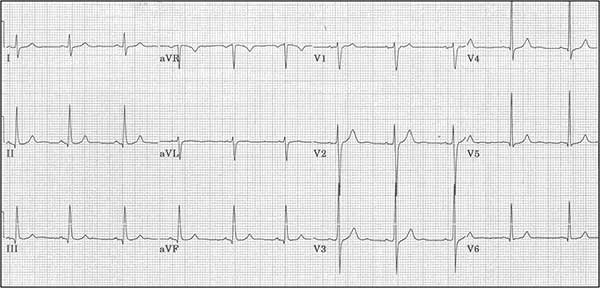
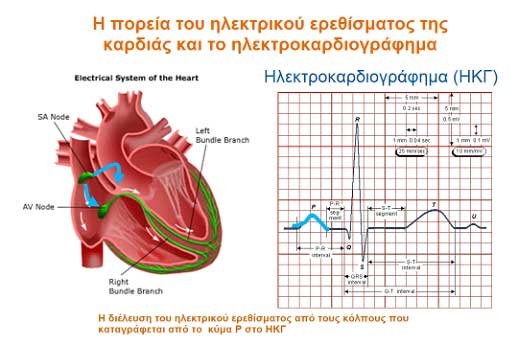
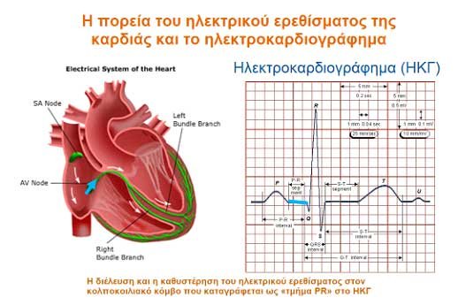
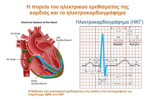
The pathway of the cardiac electrical impulse and the electrocardiogram
- The propagation of the electrical impulse through the atria, recorded as the P wave on the ECG.
- The passage and physiological delay of the electrical impulse within the atrioventricular node, recorded as the PR segment on the ECG.
- The propagation of the electrical impulse through the ventricles, recorded as the QRS complex on the ECG.