
Definition
Atrial flutter is a form of supraventricular tachycardia (SVT) characterized by an atrial rate that is significantly faster than the corresponding ventricular contraction rate. The atria typically contract at a frequency ranging from 240 to 400 beats per minute (bpm). In this setting, the atrioventricular (AV) node serves a critical gatekeeping function. It modulates conduction to prevent a 1:1 impulse transmission, which would otherwise precipitate a dangerously rapid and uncontrolled ventricular response.
Most commonly, the AV node permits the conduction of every second atrial impulse (2:1 AV conduction) or every fourth impulse (4:1 AV conduction). Consequently, an atrial rate of 260 bpm translates to a ventricular response of 130 bpm or 65 bpm, respectively. Although precise rate control is typically optimized via pharmacological agents, achieving stable ventricular rate normalization frequently poses a clinical challenge due to marked rate fluctuations, alternating between periods of severe tachycardia and profound bradycardia.
Pathophysiology / Mechanism
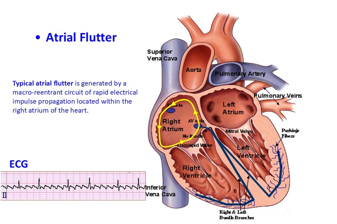
Typical atrial flutter is generated by a rapid macro-reentrant circuit (reentry loop) localized within the right atrium of the heart. This specific arrhythmia is frequently classified as isthmus-dependent. The anatomical circuit directly involves a distinct region of the right atrium known as the cavotricuspid isthmus (CTI), situated between the tricuspid valve annulus and the inferior vena cava orifice. Conversely, when the micro- or macro-reentrant circuit bypasses this anatomical isthmus and is localized within other regions of the right or left atria, the arrhythmia is classified as atypical atrial flutter. (Video)
Clinical Presentation / Symptoms
Paroxysmal or sustained episodes of atrial flutter can drive elevated ventricular rates, leading to secondary clinical manifestations such as:
- Palpitations
- Fatigue intolerance / easy fatigability
- Chest discomfort / thoracic distress
- Asthenia / generalized weakness
- Dizziness / lightheadedness
- Syncope or near-syncope
Notably, a subset of patients—particularly those in whom the ventricular rate is sufficiently controlled or "braked" by physiological or pharmacological AV nodal conduction impairment—may remain entirely asymptomatic. Atrial flutter episodes exhibit highly variable temporal patterns, lasting from a few minutes to several days or months. Patients presenting with symptomatic tachycardia require immediate clinical intervention.
Etiology
Although atrial flutter can occasionally manifest in individuals with structurally normal hearts, it predominantly develops secondary to underlying cardiovascular diseases or systemic pathological conditions. The most common etiologies and predisposing factors include:
- Prior cardiac surgery
- Heart failure (congestive heart failure)
- Coronary artery disease (CAD)
- Congenital heart disease
- Systemic arterial hypertension
- Valvular heart disease
- Right heart failure / cor pulmonale
- Pulmonary hypertension
- Chronic obstructive pulmonary disease (COPD)
Additionally, atrial flutter exhibits a high rate of coexistence with atrial fibrillation (AF), occurring concurrently in approximately 20% to 30% of the AF patient population.
Diagnosis
The definitive diagnosis of atrial flutter relies on standard surface electrocardiography (ECG) or ambulatory Holter monitoring. Procuring and preserving a high-quality ECG strip during the acute tachyarrhythmia is of paramount clinical importance, as it enables the differential diagnosis between typical (isthmus-dependent) and atypical atrial flutter. This electrophysiological differentiation is crucial when planning definitive catheter ablation therapy, given that the procedural approach, anatomical targets, and long-term prognostic outcomes vary significantly between the two entities.
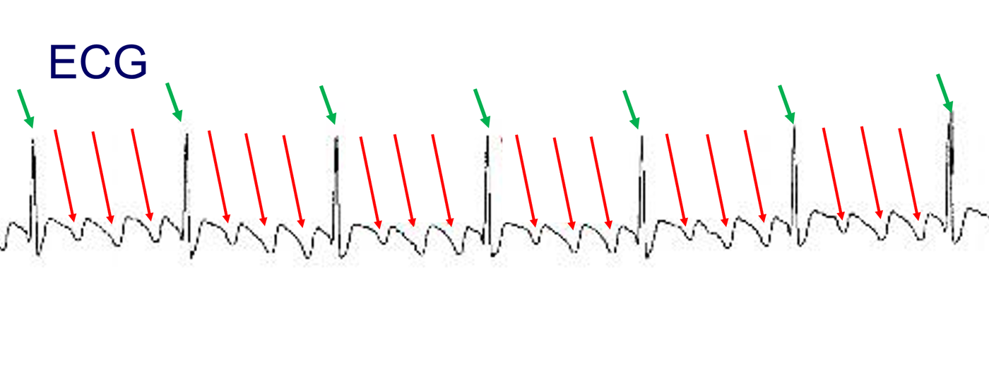 Figure 1: Surface electrocardiogram (ECG) during an active episode of atrial flutter. Red arrows delineate the rapid, regular atrial flutter waves (sawtooth pattern) occurring at a frequency of 240 bpm. Green arrows mark the corresponding regular ventricular response, controlled at a rate of 60 bpm.
Prognosis
With appropriate, guideline-directed medical management, atrial flutter is rarely a life-threatening condition. However, much like atrial fibrillation, it carries a substantially elevated risk of thromboembolic events, particularly in the presence of established clinical risk factors. These stroke risk factors include systemic hypertension, coronary or peripheral artery disease, heart failure, diabetes mellitus, advanced age, or a history of transient ischemic attack (TIA) / cerebrovascular accident (CVA). The absolute thromboembolic risk scales proportionally with the patient’s cumulative risk profile.
Current clinical data suggest that patients presenting with isolated atrial flutter may possess a marginally lower thromboembolic risk profile compared to age- and comorbidity-matched cohorts with atrial fibrillation; however, the clinical decision regarding long-term oral anticoagulation therapy must remain strictly individualized. Chronically elevated atrial frequencies can induce structural remodeling, such as atrial dilation (increased atrial volume), which is readily identifiable via transthoracic echocardiography but frequently carries no independent clinical sequelae. Conversely, if atrial flutter presents with an unmitigated, rapid ventricular response, the sustained tachycardia can induce profound myocardial dysfunction and congestive heart failure—a secondary clinical entity designated as tachycardia-induced cardiomyopathy (tachymyocardiopathy).
Therapeutic Management
Therapeutic interventions in patients diagnosed with atrial flutter are strategically directed toward three distinct clinical targets:
- Immediate termination (cardioversion) of acute symptomatic episodes.
- Optimization of ventricular rate control while the underlying arrhythmia persists (rate control).
- Permanent restoration and maintenance of normal sinus rhythm (rhythm control).
These therapeutic objectives are achieved through a combination of pharmacological therapy, electrical cardioversion, and catheter ablation.
Rate Control Management
Pharmacological agents indicated for ventricular rate mitigation via the induction of AV nodal conduction delays—while the atrial flutter remains active—primarily include beta-blockers, non-dihydropyridine calcium channel blockers (e.g., verapamil, diltiazem), and less frequently, digoxin.
Rhythm Control & Cardioversion
Class I or Class III antiarrhythmic drugs (such as amiodarone, sotalol, propafenone, or flecainide) may be utilized to suppress recurrent episodes or to attempt pharmacological conversion of an ongoing event; however, they remain largely ineffective for the definitive termination of atrial flutter. Acute rhythm restoration (termination of the arrhythmia without a guarantee against future recurrence) can be executed either pharmacologically—with intravenous ibutilide exhibiting the highest clinical efficacy—or via synchronized direct-current electrical cardioversion (DCCV) utilizing an external defibrillator.
Catheter Ablation
Catheter ablation represents the definitive first-line therapeutic strategy for virtually all patients presenting with typical atrial flutter. In high-volume, experienced electrophysiology centers, the procedural success rate approaches 100%, paired with a negligible complication rate, even when performed in elderly populations or patients with extensive systemic comorbidities.
As previously emphasized, an accurate pre-procedural ECG differentiation between typical and atypical flutter remains critical. Atypical atrial flutter frequently necessitates a more extensive, highly complex electroanatomical mapping procedure, carries a higher workload, and demonstrates less favorable long-term success rates and safety margins compared to standard cavotricuspid isthmus ablation.