
PubMed: Gerontitis D, et al. HeartRhythm Case Rep. 2026 Jun 12;12(8):878-882. e-Link
Introduction
Atrial tachycardia (AT) originating from non–pulmonary vein (PV) sources remains a significant challenge for clinical electrophysiologists. Among these sites, the superior vena cava (SVC) is 1 of the most common locations for focal triggers that can initiate or maintain atrial arrhythmias, including atrial fibrillation (AF). The specialized anatomic architecture and distinct electrophysiological properties of the cavoatrial junction are thought to provide the necessary substrate for these tachycardias, which are often characterized by high frequencies and resistance to antiarrhythmic drugs. The stan dard treatment for symptomatic SVC-AT has long been ra diofrequency (RF) catheter ablation. However, the SVC is located in a high-risk anatomic “danger zone,” flanked by the sinus node inferomedially and the right phrenic nerve posterolaterally. Thermal ablation (RF or cryoablation) carries an inherent risk of collateral damage, such as perma nent phrenic nerve palsy or sinus node dysfunction. 1–3 Pulsed field ablation (PFA) offers a tissue-selective, nonthermal alternative that can effectively isolate the SVC while sparing the adjacent phrenic nerve. However, given that sinus node cells share a similar electroporation threshold with atrial myocytes, the risk of nodal injury from extensive field delivery remains a critical consideration. This case report details the successful use of a novel balloon-in-basket PFA system in treating a complex SVC AT. To the best of our knowledge, this represents 1 of the first reported clinical cases of direct SVC-AT ablation using this specific technology, highlighting the balance between achieving electrical isolation of an arrhythmogenic vessel and preserving sinus node function.
Case report
A 71-year-old man with a history of arterial hypertension and type 2 diabetes presented with a 12-month history of paroxysmal, highly symptomatic palpitations. Previous clinical 12-lead electrocardiogram documentation during these episodes had identified a rapid, regular, narrow-QRS tachycardia with heart rates reaching 220 beats/min, characterized by P waves in the mid-R-R interval without clear polarity (Figure 1). The patient reported intense palpitations, dizziness, and presyncopal phenomena. Previous attempts at rhythm control with beta-blockers had failed; consequently, therapy was discontinued for at least 5 half-lives before the electrophysiological study to ensure unhindered arrhythmia induction.
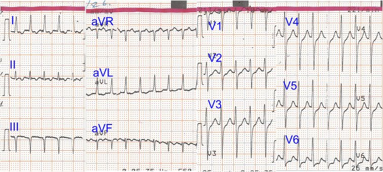
Figure 1. 12-lead electrocardiogram demonstrating the clinical atrial tachycardia with a rate of 220 beats/min.
The procedure was performed via the right femoral vein, with vascular access obtained under ultrasound guidance. A decapolar catheter (Inquiry™, Abbott, St. Paul, MN) was positioned in the coronary sinus, whereas 2 quadripolar catheters (Response™, Abbott) were placed at the His bundle position and the high right atrium or right ventricle, for recording and pacing as required. Upon admission to the electrophysiology laboratory, the patient was in sinus rhythm. However, the clinical tachycardia was easily induced after isoproterenol infusion and programmed atrial stimulation. The induced tachycardia exhibited a cycle length (CL) of 260–280 ms, with a proximal-to-distal activation sequence recorded in the coronary sinus (Figure 2). Using the EnSite X™ 3-dimensional (3D) mapping system and the Advisor™ HD Grid mapping catheter (Abbott), a high-density activation map of the right atrium was constructed. The earliest activation site was identified within the SVC, approximately 1.5–2 cm above the cavoatrial junction, suggestive of a localized reentry circuit. At this site, fragmented, high-frequency endocardial potentials were recorded, covering a significant portion of the tachycardia CL (Figure 3A). Entrainment maneuvers could not further confirm the diagnosis, given that pacing consistently resulted in tachycardia termination. Although the AT frequently degenerated into AF, necessitating electrical cardioversion, high-density mapping was successfully completed during periods of stable tachycardia, confirming the SVC’s role as the primary arrhythmic driver. During sinus rhythm, high-resolution mapping of the sinus node was performed, using the patient’s baseline heart rate as a reference. The sinus node was precisely localized at a more inferior and lateral position relative to the arrhythmic focus (Figure 3B).
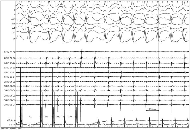
Figure 2. Tachycardia induction after programmed atrial stimulation.
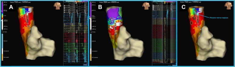
Figure 3. High-density 3-dimensional activation mapping and phrenic nerve identification. A: Activation map during tachycardia demonstrating the earliest activation site within the superior vena cava relative to the right atrium, indicative of a localized reentry pattern. Fragmented, high-frequency, and long-duration endocardial potentials are recorded at this site. B: Activation map during sinus rhythm localizing the sinus node at an inferior and lateral position relative to the arrhythmic focus. C: Phrenic nerve mapping; brown dots indicate sites of phrenic nerve capture, illustrating significant anatomic overlap with the target activation zone.
A critical step involved mapping the right phrenic nerve. High-output pacing (20 mA at 2.0 ms) along the lateral SVC wall identified a capture zone that directly overlapped with the earliest activation site (presumed localized reentry area) identified during the activation map. These points were meticulously marked on the 3D map to define the zones at risk (Figure 3C). Given the high risk of phrenic nerve injury with thermal energy, the Volt™ PFA catheter (Abbott) was selected. This 13F “balloon-in-basket” catheter was advanced via an Agilis™ NxT steerable sheath (Abbott). Before energy delivery, intravenous midazolam was administered for conscious sedation. Upon entry of the Volt PFA catheter into the SVC, the tachycardia immediately terminated owing to mechanical contact with the vessel wall (Supplemental Video 1).
PFA was then delivered using a bipolar/biphasic pulse protocol. The balloon-in-basket design ensured stable circumferential contact and allowed for complete electrical isolation of the SVC at a level superior to the mapped sinus node. A total of 3 applications were delivered to ensure sufficient overlap and achieve complete circumferential isolation. Each application was delivered using the system’s standard output (1.8 kV, biphasic), ensuring a robust and definitive isolation pattern (Figure 4, Supplemental Video 2). The delivery of pulsed energy was well tolerated by the patient, despite transient phrenic nerve capture during the applications. After the procedure, no tachycardia was inducible despite aggressive isoproterenol challenge combined with programmed atrial extrastimuli and burst pacing. Furthermore, high-output pacing after the ablation confirmed robust phrenic nerve capture, consistent with the baseline assessment. The total procedure time was 90 minutes, with a fluoroscopy time of 8.5 minutes and a total fluoroscopy dose of 85 mGy. Activated clotting time values were maintained above 300 seconds throughout the therapeutic phase of the procedure. The procedure was completed without complications, and the patient was discharged the next day in good clinical status with no recurrence of symptoms.
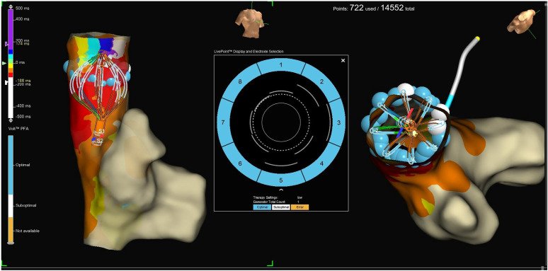
Figure 4. Pulse field ablation using the Volt catheter within the superior vena cava. The image demonstrates the balloon-in-basket catheter positioning with real-time feedback ensuring optimal tissue contact. A total of 3 overlapping pulsed energy applications were delivered to achieve complete circumferential isolation of the vessel.
Discussion
In this case, we demonstrated the successful and safe isolation of an arrhythmogenic SVC using a novel balloon-in-basket PFA system in a patient where the SVC-AT served as the primary driver for clinical symptoms. To the best of our knowledge, published clinical experience with this specific balloon-in-basket technology for isolating an arrhythmogenic SVC remains extremely limited. It is well established that the SVC is a significant site for the initiation of various arrhythmias, often acting as a trigger for AF. Several mechanisms have been implicated in SVC arrhythmogenicity, ranging from enhanced automaticity and triggered activity to various forms of reentry.4 The underlying substrate for these arrhythmias is often rooted in the complex local anatomy and the distinct electrophysiological properties of the SVC. Anatomically, the SVC contains myocardial sleeves extending up to 4–5 cm from the cavoatrial junction, with their thickness progressively decreasing cranially.5 This configuration induces conduction anisotropy, which, alongside marked conduction delays and the significant disparity in effective refractory periods between the right atrium and the SVC,6 establishes a substrate highly conducive to reentrant circuits. In the present case, the observation of fragmented potentials spanning nearly the entire tachycardia CL, combined with the reproducible induction of tachycardia via programmed atrial extrastimuli, provided strong clinical evidence of such a reentry mechanism.
In addition to the mapping findings, we observed that the baseline heart rate remained remarkably stable throughout the delivery of pulsed energy. Significant fluctuations in heart rate, including sudden acceleration or deceleration, would have been subject to various interpretations. For instance, an acceleration might have indicated the activation of the right anterior-superior atrial ganglionated plexus, but it could also have been a warning sign of impending damage to the sinus node.7 In contrast, a decrease in heart rate could have been attributed to direct sinus node injury or a transient neuromodulatory effect. The absence of these variations provided greater diagnostic clarity, underscoring the anatomic precision of the ablation. Although the tachycardia could theoretically have been managed with focal ablation at the site of slow conduction, a circumferential isolation strategy was deemed clinically superior. This approach not only mitigated the prohibitive risk of phrenic nerve injury associated with conventional RF energy but also ensured a more robust and definitive elimination of the arrhythmogenic substrate. Given that the SVC activity was proven to trigger AF, complete isolation was favored over a focal approach to prevent future clinical recurrences. This was executed rapidly and efficiently using the Volt balloon-in-basket catheter, whose design allowed for excellent apposition within the tubular anatomy of the SVC, in accordance with emerging feasibility data and recently established standardized workflows.8,9 A significant advantage of this system is the real-time feedback regarding tissue contact, ensuring that the basket is properly seated against the vessel wall before energy delivery. Furthermore, the “Sensor-Enabled” technology provided clear 3D visualization within the EnSite X map, allowing for precise orientation and the maintenance of a safe distance from the sinus node. Our experience suggests that this combination of contact indicators and advanced 3D visualization is essential for navigating the anatomic and safety challenges inherent in SVC ablation.
Addressing the broader clinical context, the identification of non-PV triggers remains a complex challenge, given that laboratory provocation with isoproterenol, adenosine, or postcardioversion observation does not always correlate with real-world clinical behavior. This was particularly evident in our patient, who presented solely with symptomatic AT and had no clinical history of AF. However, during the procedure, the SVC-originating arrhythmia was identified as a potent AF trigger, given that it repeatedly degenerated into AF. We hypothesized that although a strong trigger was present, the atrial substrate was likely not yet sufficiently remodeled to sustain AF. Thus, we opted for a targeted, trigger-based strategy, performing successful SVC isolation while deliberately refraining from prophylactic PV isolation in the absence of clinically documented AF.
Regarding safety, although PFA is widely regarded as a safe modality for preserving nerve function owing to its tissue-selective nature, recent evidence suggests that phrenic nerve injury may be more frequent than initially estimated. In particular, cases of delayed phrenic nerve palsy occurring up to 3 months after PFA have been reported.10 Nevertheless, large-scale clinical data confirm that PFA remains remarkably safe against permanent and clinically significant injury.11 In our case, despite observing phrenic nerve capture during energy delivery, postprocedural testing confirmed intact nerve function. Finally, the procedure was successfully completed under conscious sedation with midazolam, given that the Volt system’s unique pulse waveform was well tolerated, consistent with other clinical feasibility data.12 The present case highlights the potential of combining tissue-selective energy with high-density mapping to safely navigate the complex anatomy of the SVC, effectively bridging the gap between procedural precision and patient safety in the management of SVC-AT.
Conclusion
This case demonstrates that isolating an arrhythmogenic SVC using the novel balloon-in-basket PFA system is highly effective and safe. A key factor was the system’s high-density mapping capabilities, ensuring precise localization and protection of the sinus node. Excellent catheter apposition combined with real-time visualization allowed targeted energy delivery that successfully preserved adjacent critical structures, including the phrenic nerve. Furthermore, the efficiency of the procedure under conscious sedation underscores the clinical advantages of this technology. For patients where the SVC is the confirmed arrhythmic driver, this approach provides a definitive, precise, and well-tolerated therapeutic solution.
References
- Castro-Urda V, Segura-Dominguez M, Jiménez-Sánchez D, Aguilera-Agudo C, Vela-Martín P, Lorente-Ros A, García-Rodriguez D, Sánchez-Ortiz D, Pham-Trung C, García-Izquierdo E, Mingo-Santos S, Toquero-Ramos J, Fernández-Lozano I. Superior Vena Cava Isolation With Cryoballoon in AF Ablation: Randomized CAVAC AF Trial. Circ Arrhythm Electrophysiol. 2025 Jan;18(1):e012917. doi: 10.1161/CIRCEP.124.012917. Epub 2024 Dec 16. PMID: 39676679.
- Miyazaki S, Usui E, Kusa S, Taniguchi H, Ichihara N, Takagi T, Iwasawa J, Kuroi A, Nakamura H, Hachiya H, Hirao K, Iesaka Y. Prevalence and clinical outcome of phrenic nerve injury during superior vena cava isolation and circumferential pulmonary vein antrum isolation using radiofrequency energy. Am Heart J. 2014 Dec;168(6):846-53. doi: 10.1016/j.ahj.2014.09.011. Epub 2014 Sep 28. PMID: 25458647.
- Alilou S, Babaei M, Fallahtafti P, Castro-Urda V, Oraii A, Rashedi S, Di Biase L. Adjunctive superior vena cava isolation to pulmonary vein isolation in atrial fibrillation ablation: A systematic review and meta-analysis of randomized trials. Heart Rhythm. 2025 Dec 11:S1547-5271(25)03146-7. doi: 10.1016/j.hrthm.2025.12.012. Epub ahead of print. PMID: 41389862.
- Zakrzewski S, Wielusinski M, Kazmierczak J, Duda L, Hoppe W, Bazylewicz-Zakrzewska K, Kladna A, Kiedrowicz RM. Superior Vena Cava as an Arrhythmogenic Substrate in Atrial Fibrillation: Anatomical, Electrophysiological and Clinical Perspectives. J Clin Med. 2025 Oct 22;14(21):7456. doi: 10.3390/jcm14217456. PMID: 41226856; PMCID: PMC12608125.
- Kholová I, Kautzner J. Morphology of atrial myocardial extensions into human caval veins: a postmortem study in patients with and without atrial fibrillation. Circulation. 2004 Aug 3;110(5):483-8. doi: 10.1161/01.CIR.0000137117.87589.88. Epub 2004 Jul 26. PMID: 15277325.
- Fukumoto K, Takatsuki S, Kimura T, Nishiyama N, Tanimoto K, Aizawa Y, Tanimoto Y, Fukuda Y, Miyoshi S, Fukuda K. Electrophysiological properties of the superior vena cava and venoatrial junction in patients with atrial fibrillation: relevance to catheter ablation. J Cardiovasc Electrophysiol. 2014 Jan;25(1):16-22. doi: 10.1111/jce.12271. Epub 2013 Sep 19. PMID: 24103056.
- Chen G, Dong JZ, Liu XP, Zhang XY, Long DY, Sang CH, Ning M, Tang RB, Jiang CX, Ma CS. Sinus node injury as a result of superior vena cava isolation during catheter ablation for atrial fibrillation and atrial flutter. Pacing Clin Electrophysiol. 2011 Feb;34(2):163-70. doi: 10.1111/j.1540-8159.2010.02903.x. Epub 2010 Sep 30. PMID: 20883509.
- Tilz RR, Ukita K, Eitel C, Kuck KH, Hatahet S, Wenzel JP. How to isolate the superior vena cava using balloon-in-basket pulsed field ablation system: standardized workflow and initial results. Heart Rhythm O2 2026. Epub ahead of print. doi: 10.1016/j.hroo.2026.01.014.
- Papakonstantinou PE, O’Brien J. Superior vena cava isolation with 3D mapping using a novel balloon-in-basket pulsed field ablation catheter. J Interv Card Electrophysiol. 2026 Apr;69(3):327-328. doi: 10.1007/s10840-025-02158-w. Epub 2025 Oct 15. PMID: 41091364; PMCID: PMC13009074.
- Chéhirlian L, Koutbi L, Mancini J, Hourdain J, Richard-Vitton R, Wilkin M, Deharo JC, Maille B, Franceschi F. High incidence of phrenic nerve injury in patients undergoing pulsed field ablation for atrial fibrillation. Heart Rhythm. 2025 Dec;22(12):e1206-e1213. doi: 10.1016/j.hrthm.2025.08.010. Epub 2025 Aug 9. PMID: 40789463.
- Turagam MK, Aryana A, Day JD, Dukkipati SR, Hounshell T, Nair D, Natale A, Weiner S, Cheung JW, Chinitz L, Cuoco F, Daccarett M, Dandamudi S, Gambhir A, Gandhavadi M, Kim J, Metzl MD, Mikaelian B, Peress D, Romero JE, Sanchez J, Sandler DA, Shaik NA, Shehata M, Siddique SM, Singh A, Singleton MJ, Sundaram S, Vivas Y, Waks JW, Yamamura KH, Zipse M, Ahn J, Al Chekakie O, Ali M, Ascandar N, Bansal S, Beaser AD, Bisla J, Brancato S, Callans DJ, Chang-Sing P, Chothia R, Dell’Orfano J, DeLurgio DB, Doshi SK, Erickson L, Gautam S, Gottipaty V, Goyal S, Gupta S, Hajjari J, Harding JD, Hennessey J, Ho H, Ho I, Hsu JC, Huang HD, Hutchinson M, Kaplan R, Karanam S, Kaushik N, Kenigsberg DN, Khan A, Knight B, Leyton-Mange J, Lim B, Maglione TJ, Malik B, McKillop M, Mehlhorn D, Mehta D, Mittal S, Nilsson KR, Omotoye S, Oral H, Panikkath R, Patel A, Perzanowski C, Rajendra A, Razminia M, Saba S, Sanchez JM, Satti DI, Sawhney N, Sharma D, Sheppard R, Singh M, Sra J, Stone JE, Sureddi R, Taylor M, Teigeler TL, Tholakanahalli V, Trivedi A, Trulock KM, Venkataraman G, Weitz D, Williamson BD, Winters SL, Wright JM, Wu R, Yoo D, Aizer A, Alyesh DD, Amado L, Borne R, Chalfoun N, Chang D, Davis M, Ehdaie A, Gerczuk PZ, Lee JH, Michaud G, Miyama H, Mohanty S, Mora L, Mounir G, Nannapaneni N, Nazari J, Peigh G, Sauer WH, Sidney D, Varughese V, Tzou W, Gurol U, Reddy VY; MANIFEST-US Investigators. Multicenter Study on the Safety of Pulsed Field Ablation in Over 40,000 Patients: MANIFEST-US. J Am Coll Cardiol. 2026 Jan 20;87(2):172-193. doi: 10.1016/j.jacc.2025.10.051. Epub 2025 Nov 10. PMID: 41389071.
- Tilz RR, Wenzel JP, Eitel C, Popescu S, Nikorowitsch J, Kuck KH, Hatahet S. "Awake and Ablated": First experience with a balloon-in-basket PFA system without propofol. Heart Rhythm O2. 2025 Nov 21;7(2):204-211. doi: 10.1016/j.hroo.2025.11.011. PMID: 41737941; PMCID: PMC12925817.